


DR1095 Sigma-AldrichAnti-E2F2 Rabbit pAb
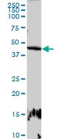

Anti-E2F2, rabbit polyclonal, recognizes the ~40-45 kDa E2F2 protein in HeLa cells and in mouse kidney. It is validated for use in Western blotting, and immunocytochemistry.
More>> Anti-E2F2, rabbit polyclonal, recognizes the ~40-45 kDa E2F2 protein in HeLa cells and in mouse kidney. It is validated for use in Western blotting, and immunocytochemistry. Less<<Anti-E2F2 Rabbit pAb MSDS (material safety data sheet) or SDS, CoA and CoQ, dossiers, brochures and other available documents.
Synonyms: Anti-E2F Transcription Factor 2
Recommended Products
Overview
Key Spec Table
| Species Reactivity | Host | Antibody Type |
|---|---|---|
| H, M | Rb | Polyclonal Antibody |
Products
| Catalogue Number | Packaging | Qty/Pack | |
|---|---|---|---|
| DR1095-100UG | 100 μg |


| Product Information | |
|---|---|
| Form | Liquid |
| Formulation | In PBS, pH 7.2. |
| Negative control | 293T cells |
| Positive control | Mouse kidney tissue, HeLa cells |
| Preservative | None |
| Quality Level | MQ100 |
| Biological Information | |
|---|---|
| Immunogen | Full-length, human E2F2 (aa 1-437) |
| Immunogen | Human |
| Host | Rabbit |
| Isotype | IgG |
| Species Reactivity |
|
| Antibody Type | Polyclonal Antibody |
| Global Trade Item Number | |
|---|---|
| Catalogue Number | GTIN |
| DR1095-100UG | 04055977226140 |