Millipore Sigma Vibrant Logo
Attention: We have moved. Merck Millipore products are no longer available for purchase on MerckMillipore.com.Learn More

05-1003 Anti-phospho-Akt (Ser473) Antibody, clone 6F5

View Products on Sigmaaldrich.com
05-1003
100 µg  
Purchase on Sigma-Aldrich

Special Offers

Overview

Replacement Information

Key Spec Table

Species ReactivityKey ApplicationsHostFormatAntibody Type
H, M, R, VrtELISA, WB, FC, IF, IHCMPurifiedMonoclonal Antibody
Description
Catalogue Number05-1003
DescriptionAnti-phospho-Akt (Ser473) Antibody, clone 6F5
Alternate Names
  • Protein kinase B
  • RAC-alpha serine/threonine-protein kinase
  • murine thymoma viral (v-akt) oncogene homolog-1
  • rac protein kinase alpha
  • v-akt murine thymoma viral oncogene homolog 1
Background InformationAkt/PKB is a Ser/Thr kinase and a major known effecter of the PI3 Kinase pathway. It is involved in multiple signaling pathways that relate to many biological processes including glucose metabolism, cell survival/apoptosis, cell cycle control, angiogenesis, differentiation, and cell growth and proliferation. In mammals three isoforms of Akt (Akt1/PKB, Akt2/PKB, and Akt3/PKB)exists. They exhibit a high degree of homology, but differ slightly in the localization of their regulatory phosphorylation sites. Akt is the predominant isoform that is in most tissues and is thought to have a dominant role in growth, survival, embryonic development, and adipocyte differentiation. Akt2 is correlated with the regulation of glucose homeostasis and is the predominant isoform expressed in insulin-responsive tissues. Akt3 is abundant in brain tissue. Each Akt isoform is composed of three functionally distinct regions: an N-terminal Pleckstrin Homology (PH) domain that provides a lipid-binding module to direct Akt to PIP3 at the cell membrane that is necessary for its activation, a central catalytic domain containing Thr308, and a C-terminal hydrophobic motif containing Ser473. The activation of Akt is dependent on a dual regulatory mechanism that requires both its translocation to the plasma membrane and dual phosphorylation on Thr308 and Ser473 by PDK1 and the TORC2 complex, respectively.
References
Product Information
FormatPurified
HS Code3002 15 90
PresentationPurified mouse monolconal IgG1κ in 0.1M Tris-Glycine (pH 7.4), 150mM NaCl with 0.05%NaN3
Quality LevelMQ100
Applications
ApplicationAnti-phospho-Akt (Ser473) Antibody, clone 6F5 detects level of phospho-Akt (Ser473) & has been published & validated for use in ELISA, WB, FC, IF, IH.
Key Applications
  • ELISA
  • Western Blotting
  • Flow Cytometry
  • Immunofluorescence
  • Immunohistochemistry
Application NotesImmunofluroescence Analysis:
EGF-treated A431 cells were fixed, permeabilized, and stained with Anti-phospho-Akt (Ser473), clone 6F5 (2 μg/mL) (red). Cy3-conjugated anti-mouse was used for detection and Phalloidin-AlexaFluor®488 (green) was used to stain actin.

Immunohistochemistry:
Anti-phospho-Akt (Ser473), clone 6F5 was diluted to 1:200, IHC-Select Detection with HRP-DAB.

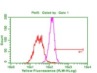
Flow Cytometry:
Anti-phospho-Akt (Ser473) was used at a 1:800 dilution for this assay.
Biological Information
ImmunogenPeptide encompassing and including phosphorylated Ser473 of human Akt1.
EpitopePhosphorylated Ser473
Clone6F5
ConcentrationPlease refer to the Certificate of Analysis for the lot-specific concentration.
HostMouse
SpecificityRecognizes Akt only when phosphorylated on Ser473. This antibody does not cross-react with Akt1/PKBα when it is not phosphorylated on Ser473.
IsotypeIgG1κ
Species Reactivity
  • Human
  • Mouse
  • Rat
  • Vertebrates
Species Reactivity NoteHuman, mouse, and rat tested. Expected to react with most species as sequence is 100% homologous with most common vertebrate.
Antibody TypeMonoclonal Antibody
Entrez Gene Number
Entrez Gene SummaryThe serine-threonine protein kinase encoded by the AKT1 gene is catalytically inactive in serum-starved primary and immortalized fibroblasts. AKT1 and the related AKT2 are activated by platelet-derived growth factor. The activation is rapid and specific, and it is abrogated by mutations in the pleckstrin homology domain of AKT1. It was shown that the activation occurs through phosphatidylinositol 3-kinase. In the developing nervous system AKT is a critical mediator of growth factor-induced neuronal survival. Survival factors can suppress apoptosis in a transcription-independent manner by activating the serine/threonine kinase AKT1, which then phosphorylates and inactivates components of the apoptotic machinery. Multiple alternatively spliced transcript variants have been found for this gene. [provided by RefSeq].
Gene Symbol
  • AKT
  • C-AKT
  • EC 2.7.11.1
  • MGC99656
  • MGC99656
  • PKB-ALPHA
  • PRKBA
  • RAC
  • RAC-ALPHA
  • RAC-PK-alpha
Modifications
  • Phosphorylation
Purification MethodProtein G Purified
UniProt Number
UniProt SummaryFUNCTION: General protein kinase capable of phosphorylating several known proteins. Phosphorylates TBC1D4. Signals downstream of phosphatidylinositol 3-kinase (PI(3)K) to mediate the effects of various growth factors such as platelet-derived growth factor (PDGF), epidermal growth factor (EGF), insulin and insulin-like growth factor I (IGF-I). Plays a role in glucose transport by mediating insulin-induced translocation of the GLUT4 glucose transporter to the cell surface. Mediates the antiapoptotic effects of IGF-I. Mediates insulin-stimulated protein synthesis, partly by playing a role in both insulin-induced phosphorylation of 4E-BP1 and in insulin-induced activation of p70 S6 kinase. Promotes glycogen synthesis by mediating the insulin-induced activation of glycogen synthase. Ref.4 Ref.8 Ref.12 Ref.13 Ref.16 Ref.20

Catalytic activity ATP + a protein = ADP + a phosphoprotein. Ref.4 Ref.20 Ref.1

ENZYME REGULATION:Three specific sites, one in the kinase domain (Thr-308) and the two other ones in the C-terminal regulatory region (Ser-473 and Tyr-474), need to be phosphorylated for its full activation. Ref.8 Ref.6

SUBUNIT STRUCTURE: Interacts with AGAP2 isoform 2 (PIKE-A) in the presence of guanine nucleotides. The C-terminus interacts with CCDC88A/GRDN and THEM4. Interacts with AKTIP. Interacts (via PH domain) with MTCP1, TCL1A AND TCL1B. Interacts with CDKN1B; the interaction phosphorylates CDKN1B promoting 14-3-3 binding and cell-cycle progression. Ref.13 Ref.16 Ref.20 Ref.9 Ref.10 Ref.11 Ref.15 Ref.17 Ref.18 Ref.19

SUBCELLULAR LOCATION: Cytoplasm. Nucleus. Cell membrane. Note= Nucleus after activation by integrin-linked protein kinase 1 (ILK1). Nuclear translocation is enhanced by interaction with TCL1A. Ref.11

TISSUE SPECIFICTY: In all human cell types so far analyzed. Ref.4

DOMAIN: Binding of the PH domain to the phosphatidylinositol 3-kinase alpha (PI(3)K) results in its targeting to the plasma membrane.

The AGC-kinase C-terminal mediates interaction with THEM4.

PTM: Phosphorylation on Thr-308, Ser-473 and Tyr-474 is required for full activity. Ser-473 phosphorylation by the Rictor-mTor complex favors Thr-308 phosphorylation by PDPK1. Ser-473 phosphorylation is enhanced by interaction with AGAP2 isoform 2 (PIKE-A). Ser-473 phosphorylation is enhanced in focal cortical dysplasias with Taylor-type balloon cells.

SIMILARITY: Belongs to the protein kinase superfamily. AGC Ser/Thr protein kinase family. RAC subfamily.

Contains 1 AGC-kinase C-terminal domain.

Contains 1 PH domain.

Contains 1 protein kinase domain.
Molecular Weight60 kDa
Physicochemical Information
Dimensions
Materials Information
Toxicological Information
Safety Information according to GHS
Safety Information
Product Usage Statements
Quality AssuranceWestern Blot Analysis:
0.5-1 μg/mL of this lot detected phosphorylated Akt in lysates from mouse NIH-3T3 fibroblasts treated with 100 ng/mL PDGF for 20 minutes.
Usage Statement
  • Unless otherwise stated in our catalog or other company documentation accompanying the product(s), our products are intended for research use only and are not to be used for any other purpose, which includes but is not limited to, unauthorized commercial uses, in vitro diagnostic uses, ex vivo or in vivo therapeutic uses or any type of consumption or application to humans or animals.
Storage and Shipping Information
Storage ConditionsStable for 1 year at 2-8ºC from date of receipt.
Packaging Information
Material Size100 µg
Transport Information
Supplemental Information
Specifications
Global Trade Item Number
Catalogue Number GTIN
05-1003 04053252329494

Documentation

Anti-phospho-Akt (Ser473) Antibody, clone 6F5 MSDS

Title

Safety Data Sheet (SDS) 

Anti-phospho-Akt (Ser473) Antibody, clone 6F5 Certificates of Analysis

TitleLot Number
Anti-phospho-Akt (Ser473), clone 6F5 - 2384001 2384001
Anti-phospho-Akt (Ser473), clone 6F5 - 2430475 2430475
Anti-phospho-Akt (Ser473), clone 6F5 - 1975108 1975108
Anti-phospho-Akt (Ser473), clone 6F5 - 1998946 1998946
Anti-phospho-Akt (Ser473), clone 6F5 - 2061572 2061572
Anti-phospho-Akt (Ser473), clone 6F5 - 2161212 2161212
Anti-phospho-Akt (Ser473), clone 6F5 - 2224460 2224460
Anti-phospho-Akt (Ser473), clone 6F5 - 2298421 2298421
Anti-phospho-Akt (Ser473), clone 6F5 - 2476994 2476994
Anti-phospho-Akt (Ser473), clone 6F5 - 3288731 3288731

References

Reference overviewPub Med ID
3-Phosphoinositide-dependent protein kinase 1 (PDK1) phosphorylates and activates the p70 S6 kinase in vivo and in vitro.
Alessi, D R, et al.
Curr. Biol., 8: 69-81 (1998)  1998

9427642 9427642
PDK1, one of the missing links in insulin signal transduction?
Cohen, P, et al.
FEBS Lett., 410: 3-10 (1997)  1997

Show Abstract
9247112 9247112
3-Phosphoinositide-dependent protein kinase-1 (PDK1): structural and functional homology with the Drosophila DSTPK61 kinase.
Alessi, D R, et al.
Curr. Biol., 7: 776-89 (1997)  1997

9368760 9368760

Related Products & Applications

Related Products

Product Families

Categories

Life Science Research > Antibodies and Assays > Primary Antibodies