Millipore Sigma Vibrant Logo
Attention: We have moved. Merck Millipore products are no longer available for purchase on MerckMillipore.com.Learn More

MABS1916 Anti-c-Met Antibody, clone seeMet 13

View Products on Sigmaaldrich.com
MABS1916
100 µL  
Purchase on Sigma-Aldrich

Special Offers

Overview

Replacement Information

Key Spec Table

Species ReactivityKey ApplicationsHostFormatAntibody Type
HIP, ICC, FC, InhibitionMPurifiedMonoclonal Antibody
Description
Catalogue NumberMABS1916
DescriptionAnti-c-Met Antibody, clone seeMet 13
Alternate Names
  • Hepatocyte growth factor receptor
  • HGF receptor
  • HGF/SF receptor
  • Proto-oncogene c-Met
  • Scatter factor receptor
  • SF receptor
  • Tyrosine-protein kinase Met
Background InformationHepatocyte growth factor receptor (EC 2.7.10.1; UniProt P08581; also known as HGF receptor, HGF/SF receptor, Proto-oncogene c-Met, Scatter factor receptor, SF receptor, Tyrosine-protein kinase Met) is encoded by the MET (also known as AUTS9, HCC, RCCP) gene (Gene ID 4233) in human. HGF receptor or c-Met is initially produced as a 1390-a.a. precursor protein with an N-terminal signal peptide, postranslational proteolytic cleavage yields the mature tyrosine kinase receptor composed of an extracellular alpha chain (a.a. 25-307) linked via a disulfide bond to a transmembrane beta chain with an extracellular domain (a.a. 308-932), transmembrane helix (a.a. 933-955), and a cytoplasmic domain (a.a. 956-1390). The alpha-chain and the extracellular portion of beta-chain form the Sema domain that mediates receptor dimerization and ligand binding. The cytoplasmic portion of beta-chain contains the kinase domain (a.a. 1078-1345) and a C-terminal tail that contains a docking region (a.a. 1212-1390) for effector and adapter proteins essential for downstream signaling. Hepatocyte growth factor (HGF) is the only known c-Met ligand. HGF binding induces c-Met dimerization and autophosphorylation on Y1230, Y1234 and Y1235, leading to a conformation change and further phosphorylation on Y1349 and Y1356, making C-terminal tail docking region available for adaptor and signalling molecules. c-Met mediates a wide-range of biological activities, including cell proliferation, motility, angiogenesis and morphogenesis, and is an attractive target for cancer therapy. Aberrant c-Met activation/signalling promotes cancer cell proliferation and is associated with poor prognosis in various human cancers. Upregulated c-Met expression is also known to cause resistance against HER2, EGFR and B-RAF inhibitory drugs in cancer treatment.
References
Product Information
FormatPurified
PresentationPurified mouse IgG1 in PBS without preservatives.
Quality LevelMQ100
Applications
Application
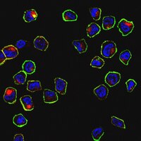
Detect Hepatocyte growth factor receptor using this mouse monoclonal Anti-c-Met, clone seeMet 13, Cat. No. MABS1916, validated for use in Flow Cytometry, Immunocytochemistry, Immunoprecipitation, and Inhibition studies.
Key Applications
  • Immunoprecipitation
  • Immunocytochemistry
  • Flow Cytometry
  • Inhibition
Application NotesFlow Cytometry Analysis: 0.2 µL from a representative lot detected c-Met immunoreactivity on the surface of one million SNU-5 cells.

Flow Cytometry Analysis: A representative lot immunostained live SNU-5 cells. A slight decrease in antibody immunoreactivity was observed when the temperature was dropped from 37°C to 4°C (Wong, J.S., et al. (2013). Oncotarget. 4(7):1019-1036).

Immunocytochemistry Analysis: A representative lot immunostained live SNU-5 cells. Indirect fluorescence labelling following subsequent cell fixation and permeabilization revealed increased antibody cytoplasmic internalization when antibody incubation was performed at 37°C than at 4°C (Wong, J.S., et al. (2013). Oncotarget. 4(7):1019-1036).

Immunoprecipitation Analysis: A representative lot immunoprecipitated c-Met from SNU-5 cell lysate (Wong, J.S., et al. (2013). Oncotarget. 4(7):1019-1036).

Inhibition Analysis: A representative lot prevented HGF-induced scatter of HaCaT cells. Clone seeMet 13 inhibited cell proliferation by blocking cell division without inducing apoptosis (Wong, J.S., et al. (2013). Oncotarget. 4(7):1019-1036).

Note: seeMet 13 exhibited low reactivity and specificity towards denatured c-Met by Western blotting. This monoclonal antibody is not recommended for Western blotting application (Wong, J.S., et al. (2013). Oncotarget. 4(7):1019-1036).
Biological Information
ImmunogenHis-tagged recombinant human c-Met alpha chain (Wong, J.S., et al. (2013). Oncotarget. 4(7):1019-1036).
CloneseeMet 13
ConcentrationPlease refer to lot specific datasheet.
HostMouse
SpecificityClone seeMet 13 bound strongly to native c-Met by flow cytometry, while exhibiting low reactivity and specificity towards denatured c-Met by Western blotting (Wong, J.S., et al. (2013). Oncotarget. 4(7):1019-1036).
IsotypeIgG1κ
Species Reactivity
  • Human
Species Reactivity NoteHuman.
Antibody TypeMonoclonal Antibody
Entrez Gene Number
Gene Symbol
  • MET
  • AUTS9
  • HCC
  • RCCP
Purification MethodProtein G purified.
UniProt Number
Molecular Weight32.51 kDa (alpha chain; a.a. 25-307), 155.5/153.0 kDa (pro-/mature uncleaved isoform 1), 157.7/155.2 kDa (pro-/mature uncleaved isoform 2), 85.75/83.25 kDa (pro-/mature uncleaved isoform 3) calculated.
Physicochemical Information
Dimensions
Materials Information
Toxicological Information
Safety Information according to GHS
Safety Information
Product Usage Statements
Quality AssuranceEvaluated by Immunocytochemistry in SNU-5 cells.

Immunocytochemistry Analysis: A 1:500 dilution of this antibody detected both surface and internalized c-Met by fluorescent immunocytochemistry staining of 4% paraformaldehyde-fixed, 0.3% Triton X-100-permeabilized SNU-5 cells.
Usage Statement
  • Unless otherwise stated in our catalog or other company documentation accompanying the product(s), our products are intended for research use only and are not to be used for any other purpose, which includes but is not limited to, unauthorized commercial uses, in vitro diagnostic uses, ex vivo or in vivo therapeutic uses or any type of consumption or application to humans or animals.
Storage and Shipping Information
Storage ConditionsStable for 1 year at -20°C from date of receipt.

Handling Recommendations: Upon receipt and prior to removing the cap, centrifuge the vial and gently mix the solution. Aliquot into microcentrifuge tubes and store at -20°C. Avoid repeated freeze/thaw cycles, which may damage IgG and affect product performance.
Packaging Information
Material Size100 µL
Transport Information
Supplemental Information
Specifications
Global Trade Item Number
Catalogue Number GTIN
MABS1916 04054839090233