Millipore Sigma Vibrant Logo
Attention: We have moved. Merck Millipore products are no longer available for purchase on MerckMillipore.com.Learn More

IM32 Anti-TIMP-1 (Ab-1) Mouse mAb (7-6C1)

Overview

Replacement Information

Key Spec Table

Species ReactivityHostAntibody Type
B, H, RMMonoclonal Antibody

Products

Catalogue NumberPackaging Qty/Pack
IM32-100UGCN 塑膠安瓿;塑膠針藥瓶 100 μg
Description
OverviewRecognizes the ~28.5 kDa TIMP-1 protein. Also recognizes MMP/TIMP-1 complexes.
Catalogue NumberIM32
Brand Family Calbiochem®
SynonymsAnti-Tissue Inhibitor of Metalloproteinase-1
Application Data
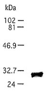
Detection of TIMP-1 by immunoblotting. Sample: Whole cell lysate from CHO cells. Primary antibody: Anti-TIMP-1 (Ab-1) Mouse mAb (7-6C1) (Cat. No. IM32) (1 µg/ml). Detection: chemiluminescence.
References
ReferencesFujimoto, N. and Iwata, K., 1994. Connective Tissue 26, 237.
Cottam, D. W. and Rees, R. C., 1993. Intl. J. Oncol. 2, 861.
Stetler-Stevenson, W. G., et al. 1993. FASEB J. 7, 1434.
Woessner, J. F., 1991. FASEB J. 5, 2145.
Liotta, L. A. and Stetler-Stevenson, W. G. 1990. in Seminars in Cancer Biology ed. M. M. Gottesman. Vol. 99-106.
Product Information
DeclarationManufactured by Daiichi Fine Chemical Co., Ltd. Not available for sale in Japan.
FormLiquid
FormulationIn 100 mM sodium phosphate buffer, 0.1% BSA, pH 7.0.
Positive controlrTIMP-1 Protein (Cat. Nos. PF019 or PF020)
Preservative≤0.1% sodium azide
Quality LevelMQ100
Applications
Application ReferencesImmunoblotting Kodama, S., et al. 1990. J. Immunol. Meth. 127, 103. Imunohistochemistry Okada, Y., et al. 1990. Virchows Arch. B Cell Pathol. 59, 305. Paraffin Sections Romanic, A.M., et al. 1998. Stroke 29, 1020. Immunocytochemistry Zhao, W.Q., et al. 1998. J. Cell Sci. 111, 1147. Original Clone Kodama, S., et al. 1987. Collagen Rel. Res. 7, 341.
Key Applications Immunoblotting (Western Blotting)
Immunocytochemistry
Paraffin Sections
Application NotesImmunoblotting (1 µg/ml, see application references)
Immunocytochemistry (see application references)
Paraffin Sections (25 µg/ml, see application references, see comments)
Application CommentsAlso recognizes TIMP-1/MMP complexes.This antibody has been used to stain paraffin sections fixed with PLP (perioddate-lysine-4% paraformaldehyde) and 10% formaldehyde (using 25 µg/ml antibody). Antibody should be titrated for optimal results in individual systems.
Biological Information
Immunogenpurified, bovine dental pulp TIMP-1
ImmunogenBovine
Clone7-6C1
HostMouse
IsotypeIgG₁
Species Reactivity
  • Bovine
  • Human
  • Rat
Antibody TypeMonoclonal Antibody
Concentration Label Please refer to vial label for lot-specific concentration
Physicochemical Information
Dimensions
Materials Information
Toxicological Information
Safety Information according to GHS
Safety Information
Product Usage Statements
Storage and Shipping Information
Ship Code Blue Ice Only
Toxicity Standard Handling
Storage -20°C
Avoid freeze/thaw Avoid freeze/thaw
Do not freeze Ok to freeze
Special InstructionsFollowing initial thaw, aliquot and freeze (-20°C).
Packaging Information
Transport Information
Supplemental Information
Specifications
Global Trade Item Number
Catalogue Number GTIN
IM32-100UGCN 07790788053765

Documentation

Anti-TIMP-1 (Ab-1) Mouse mAb (7-6C1) MSDS

Title

Safety Data Sheet (SDS) 

Anti-TIMP-1 (Ab-1) Mouse mAb (7-6C1) Certificates of Analysis

TitleLot Number
IM32

References

Reference overview
Fujimoto, N. and Iwata, K., 1994. Connective Tissue 26, 237.
Cottam, D. W. and Rees, R. C., 1993. Intl. J. Oncol. 2, 861.
Stetler-Stevenson, W. G., et al. 1993. FASEB J. 7, 1434.
Woessner, J. F., 1991. FASEB J. 5, 2145.
Liotta, L. A. and Stetler-Stevenson, W. G. 1990. in Seminars in Cancer Biology ed. M. M. Gottesman. Vol. 99-106.
Data Sheet

Note that this data sheet is not lot-specific and is representative of the current specifications for this product. Please consult the vial label and the certificate of analysis for information on specific lots. Also note that shipping conditions may differ from storage conditions.

Revision27-August-2007 RFH
SynonymsAnti-Tissue Inhibitor of Metalloproteinase-1
ApplicationImmunoblotting (1 µg/ml, see application references)
Immunocytochemistry (see application references)
Paraffin Sections (25 µg/ml, see application references, see comments)
Application Data
Detection of TIMP-1 by immunoblotting. Sample: Whole cell lysate from CHO cells. Primary antibody: Anti-TIMP-1 (Ab-1) Mouse mAb (7-6C1) (Cat. No. IM32) (1 µg/ml). Detection: chemiluminescence.
DescriptionPurified mouse monoclonal antibody (see application references). Recognizes the ~28 kDa TIMP-1 protein.
BackgroundMatrix metalloproteinases (MMPs) are a family of enzymes that are responsible for the degradation of extracellular matrix components such as collagen, laminin and proteoglycans. In addition to sequence homology, all MMPs share the following characteristics: the catalytic mechanism is dependent upon a zinc ion at the active center, they cleave one or more extracellular matrix components, they are secreted as zymogens which are activated by removal of an approximately 10 kDa segment from the N-terminus and their activity is regulated by endogenous inhibitors. These enzymes are involved in normal physiological processes such as embryogenesis and tissue remodeling and may play an important role in arthritis, periodontitis, and metastasis. The activation and activity of MMPs are regulated by a family of endogenous inhibitors, tissue inhibitors of metalloproteinase (TIMP). TIMP-1 (also called EPA, Fibroblast collagenase inhibitor or collagenase inhibitor) is a 28 kDa glycoprotein that is expressed by a variety of cell types. It forms a non-covalent, stoichiometric complex with both latent and active MMPs. TIMP-1 preferentially binds and inhibits MMP-9. TIMPs are capable of altering the metastatic potential of cancer cells and have been shown to inhibit invasion and metastasis in animal models.
HostMouse
Immunogen speciesBovine
Immunogenpurified, bovine dental pulp TIMP-1
Clone7-6C1
IsotypeIgG₁
Speciesbovine, human, not mouse, rat
Positive controlrTIMP-1 Protein (Cat. Nos. PF019 or PF020)
FormLiquid
FormulationIn 100 mM sodium phosphate buffer, 0.1% BSA, pH 7.0.
Concentration Label Please refer to vial label for lot-specific concentration
Preservative≤0.1% sodium azide
CommentsAlso recognizes TIMP-1/MMP complexes.This antibody has been used to stain paraffin sections fixed with PLP (perioddate-lysine-4% paraformaldehyde) and 10% formaldehyde (using 25 µg/ml antibody). Antibody should be titrated for optimal results in individual systems.
Storage Avoid freeze/thaw
-20°C
Do Not Freeze Ok to freeze
Special InstructionsFollowing initial thaw, aliquot and freeze (-20°C).
Toxicity Standard Handling
ReferencesFujimoto, N. and Iwata, K., 1994. Connective Tissue 26, 237.
Cottam, D. W. and Rees, R. C., 1993. Intl. J. Oncol. 2, 861.
Stetler-Stevenson, W. G., et al. 1993. FASEB J. 7, 1434.
Woessner, J. F., 1991. FASEB J. 5, 2145.
Liotta, L. A. and Stetler-Stevenson, W. G. 1990. in Seminars in Cancer Biology ed. M. M. Gottesman. Vol. 99-106.
Application referencesImmunoblotting Kodama, S., et al. 1990. J. Immunol. Meth. 127, 103. Imunohistochemistry Okada, Y., et al. 1990. Virchows Arch. B Cell Pathol. 59, 305. Paraffin Sections Romanic, A.M., et al. 1998. Stroke 29, 1020. Immunocytochemistry Zhao, W.Q., et al. 1998. J. Cell Sci. 111, 1147. Original Clone Kodama, S., et al. 1987. Collagen Rel. Res. 7, 341.