Millipore Sigma Vibrant Logo
Attention: We have moved. Merck Millipore products are no longer available for purchase on MerckMillipore.com.Learn More

AM76 Anti-TBK1 Mouse mAb (108A429)

Overview

Replacement Information

Key Spec Table

Species ReactivityHostAntibody Type
HMMonoclonal Antibody

Products

Catalogue NumberPackaging Qty/Pack
AM76-100UGCN 塑膠安瓿;塑膠針藥瓶 100 μg
Description
OverviewRecognizes the ~80 kDa TBK1 protein in HEK293 cells.
Catalogue NumberAM76
Brand Family Calbiochem®
Application Data
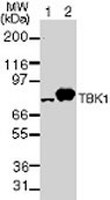
Detection of human TBK1 by immunoblotting. Samples: Whole cell lysates from 293 cells untransfected (lane 1) and transfected with human TBK1 cDNA (lane 2). Primary antibody: Anti-TBK1 Mouse mAb (108A429) (Cat. No. AM76) (2 µg/ml). Detection: chemiluminescence.

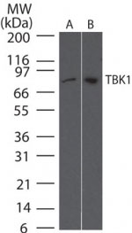
Detection of TBK1 by immunoblotting. Samples: Whole cell lysates from human Daudi cells (lane 1) and mouse RAW cells (lane 2). Primary antibody: Anti-TBK1 Mouse mAb (108A429) (Cat. No. AM76) (2 µg/ml). Detection: chemiluminescence.
References
ReferencesPomeerantz, J.L. and Baltimore, D. 1999. EMBO J. 18, 6694.
Didonato, J.A., et al. 1997. Nature 388, 548.
Lee, F.S., et al. 1997. Cell 88, 213.
Malinin, N.L., et al. 1997. Nature 385, 540.
Mercurio, F., et al. 1997. Science 278, 860.
Regnier, C.H., et al. 1997. Cell 90, 373.
Verma, I. and Stevenson, J.K. 1997. Proc. Natl. Acad. Sci. USA 94, 11758.
Verma, I.M., et al. 1995. Genes Dev. 9, 2723.
Product Information
FormLiquid
FormulationIn PBS, 0.2% gelatin.
Negative controlNormal mouse serum
Positive controlHEK293 cells
Preservative≤0.1% sodium azide
Quality LevelMQ100
Applications
Key Applications Immunoblotting (Western Blotting)
Application NotesImmunoblotting (2 µg/ml)
Application CommentsAntibody should be titrated for optimal results in an individual systems.
Biological Information
Immunogenfull-length, recombinant, human TBK1 fused to a His•Tag® sequence
ImmunogenHuman
Clone108A429
HostMouse
IsotypeIgG₁
Species Reactivity
  • Human
Antibody TypeMonoclonal Antibody
Concentration Label Please refer to vial label for lot-specific concentration
Physicochemical Information
Dimensions
Materials Information
Toxicological Information
Safety Information according to GHS
Safety Information
Product Usage Statements
Storage and Shipping Information
Ship Code Blue Ice Only
Toxicity Standard Handling
Storage -20°C
Avoid freeze/thaw Avoid freeze/thaw
Do not freeze Ok to freeze
Special InstructionsFollowing initial thaw, aliquot and freeze (-20°C).
Packaging Information
Transport Information
Supplemental Information
Specifications
Global Trade Item Number
Catalogue Number GTIN
AM76-100UGCN 04055977228045

Documentation

Anti-TBK1 Mouse mAb (108A429) MSDS

Title

Safety Data Sheet (SDS) 

Anti-TBK1 Mouse mAb (108A429) Certificates of Analysis

TitleLot Number
AM76

References

Reference overview
Pomeerantz, J.L. and Baltimore, D. 1999. EMBO J. 18, 6694.
Didonato, J.A., et al. 1997. Nature 388, 548.
Lee, F.S., et al. 1997. Cell 88, 213.
Malinin, N.L., et al. 1997. Nature 385, 540.
Mercurio, F., et al. 1997. Science 278, 860.
Regnier, C.H., et al. 1997. Cell 90, 373.
Verma, I. and Stevenson, J.K. 1997. Proc. Natl. Acad. Sci. USA 94, 11758.
Verma, I.M., et al. 1995. Genes Dev. 9, 2723.
Data Sheet

Note that this data sheet is not lot-specific and is representative of the current specifications for this product. Please consult the vial label and the certificate of analysis for information on specific lots. Also note that shipping conditions may differ from storage conditions.

Revision17-August-2007 RFH
ApplicationImmunoblotting (2 µg/ml)
Application Data
Detection of human TBK1 by immunoblotting. Samples: Whole cell lysates from 293 cells untransfected (lane 1) and transfected with human TBK1 cDNA (lane 2). Primary antibody: Anti-TBK1 Mouse mAb (108A429) (Cat. No. AM76) (2 µg/ml). Detection: chemiluminescence.

Detection of TBK1 by immunoblotting. Samples: Whole cell lysates from human Daudi cells (lane 1) and mouse RAW cells (lane 2). Primary antibody: Anti-TBK1 Mouse mAb (108A429) (Cat. No. AM76) (2 µg/ml). Detection: chemiluminescence.
DescriptionProtein G purified mouse monoclonal antibody. Recognizes the ~80 kDa TBK1 protein.
BackgroundNuclear factor κB (NF-κB) is sequestered in cell cytoplasm by the IκB family of inhibitory proteins that mask the nuclear localization signal of NF-κB, preventing its translocation to the nucleus. Stimulus by tumor necrosis factor or other cytokines results in the phosphorylation and degradation of IκB, releasing NF-κB dimers. These dimers then translocate to the nucleus and activate target genes. IκBα synthesis is autoregulated. IκB proteins are phosphorylated by an IκB kinase made up of at least three proteins; IKK1/α, IKK2/β, and IKK3/γ. IKK 1/α and IKK2/β are phosphorylated by NF-κB-inducing kinase (NIK) and MAP kinase kinase kinase-1 (MEKK1) respectively. TANK-binding kinase (TBK1) has recently been identified as a member of the IKK family. TRAF-associated NF-κB activator (TANK) interacts with TBK1 and activates NF-κB in a kinase-dependent manner.
HostMouse
Immunogen speciesHuman
Immunogenfull-length, recombinant, human TBK1 fused to a His•Tag® sequence
Clone108A429
IsotypeIgG₁
Specieshuman
Positive controlHEK293 cells
Negative controlNormal mouse serum
FormLiquid
FormulationIn PBS, 0.2% gelatin.
Concentration Label Please refer to vial label for lot-specific concentration
Preservative≤0.1% sodium azide
CommentsAntibody should be titrated for optimal results in an individual systems.
Storage Avoid freeze/thaw
-20°C
Do Not Freeze Ok to freeze
Special InstructionsFollowing initial thaw, aliquot and freeze (-20°C).
Toxicity Standard Handling
ReferencesPomeerantz, J.L. and Baltimore, D. 1999. EMBO J. 18, 6694.
Didonato, J.A., et al. 1997. Nature 388, 548.
Lee, F.S., et al. 1997. Cell 88, 213.
Malinin, N.L., et al. 1997. Nature 385, 540.
Mercurio, F., et al. 1997. Science 278, 860.
Regnier, C.H., et al. 1997. Cell 90, 373.
Verma, I. and Stevenson, J.K. 1997. Proc. Natl. Acad. Sci. USA 94, 11758.
Verma, I.M., et al. 1995. Genes Dev. 9, 2723.