Millipore Sigma Vibrant Logo
Attention: We have moved. Merck Millipore products are no longer available for purchase on MerckMillipore.com.Learn More

06-1433 Anti-phospho-FGFR-1 (Tyr653/Tyr654) Antibody

View Products on Sigmaaldrich.com
06-1433
100 µg  
Purchase on Sigma-Aldrich

Special Offers

Overview

Replacement Information

Key Spec Table

Species ReactivityKey ApplicationsHostFormatAntibody Type
H, M, R, Ch, B, XnWB, PIARbAffinity PurifiedPolyclonal Antibody
Description
Catalogue Number06-1433
DescriptionAnti-phospho-FGFR-1 (Tyr653/Tyr654) Antibody
Alternate Names
  • Fibroblast growth factor receptor 1
  • EC:2.7.10.1
  • FGFR-1
  • Basic fibroblast growth factor receptor 1
  • BFGFR
  • bFGF-R-1
  • Fms-like tyrosine kinase 2
  • FLT-2
  • N-sam
  • Proto-oncogene c-Fgr
  • CD331
Background InformationFibroblast growth factor receptor 1 (UniProt: P11362; also known as EC:2.7.10.1, FGFR-1, Basic fibroblast growth factor receptor 1, BFGFR, bFGF-R-1, Fms-like tyrosine kinase 2, FLT-2, N-sam, Proto-oncogene c-Fgr, CD331) is encoded by the FGFR1 (also known as BFGFR, CEK, FGFBR, FLG, FLT2, HBGFR) gene (Gene ID: 2260) in human. FGF-R1 is a single-pass type I membrane glycoprotein with serves as a receptor for fibroblast growth factor and plays a role in the regulation of embryonic development, cell proliferation, differentiation and migration. It is required for normal mesoderm patterning and correct axial organization during embryonic development, normal skeletogenesis and normal development of the gonadotropin-releasing hormone (GnRH) neuronal system. FGFR-1 is synthesized with a signal peptide (aa 1-21), which is subsequently cleaved off to generate the mature form that contains an extracellular domain (aa 22-376), a transmembrane domain (aa 377-397), and a cytoplasmic domain (aa 398-822). FGF-R1 undergoes autophosphorylation and upon binding of FGF family members together with heparan sulfate proteoglycan or heparin promotes receptor dimerization and autophosphorylation on tyrosine residues. Initial autophosphorylation at tyrosine 653 increases the kinase activity by a factor of 50 to 100. Following this initial phosphorylation tyrosine 583 is phosphorylated followed by phosphorylation of tyrosines 463, 766, 583, and 585. In a third stage, tyrosine 654 is autophosphorylated, resulting in a further tenfold increase its kinase activity. Phosphotyrosine residues provide docking sites for interacting proteins and so are crucial for FGF-R1 function and its regulation. The downstream targets of PGF-R1 include phospholipase C g1 (PLCg1) FGF receptor substrate 2, GRB2-associated-binding protein 1 (GAB1), and SH2 domain-containing adapter protein B (SHB2). This ZooMAb® recombinant monoclonal antibody, generated by our propriety technology, offers significantly enhanced specificity, affinity, reproducibility, and stability over conventional monoclonals.
References
Product Information
FormatAffinity Purified
Control
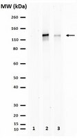
  • Untreated and lambda phosphatase-treated HEK293T cells transfected with FGFR-1
PresentationPurified rabbit polyclonal antibody in buffer containing 0.1 M Tris-Glycine (pH 7.4), 150 mM NaCl with 0.05% sodium azide.
Quality LevelMQ100
Applications
ApplicationAnti-phospho-FGFR-1 (Tyr653/Tyr654), Cat. No. 06-1433, is a Rabbit polyclonal antibody that detects basic fibroblast growth factor receptor 1 phosphorylated on Tyr 653/654 and is tested for use in Peptide Inhibition Assay and use in Western Blotting.
Key Applications
  • Western Blotting
  • Peptide Inhibition Assay
Application NotesPeptide Inhibition Analysis: A 1:5,000 dilution from a representative lot failed to detect the target band in lysate from HEK293 cells transfected with human FGFR1 when preblocked with the immunogen phosphopeptide, but not with a corresponding non-phosphopeptide.

Note: Actual optimal working dilutions must be determined by end user as specimens, and experimental conditions may vary with the end user
Biological Information
ImmunogenKLH-conjugated linear peptide corresponding to 12 amino acids surrounding phosphotyrosine 653/654 from the C-terminal, cytoplasmic domain of human Fibroblast growth factor receptor 1 (FGFR1).
EpitopePhosphorylated Tyr653/Tyr654
ConcentrationPlease refer to the Certificate of Analysis for the lot-specific concentration.
HostRabbit
SpecificityThis rabbit polyclonal antibody specifically detects Fibroblast growth factor receptor 1 (FGFR1) phosphorylated on tyrosine 653/654.
Species Reactivity
  • Human
  • Mouse
  • Rat
  • Chicken
  • Bovine
  • Xenopus
Species Reactivity NoteHuman. Predicted to react with Bovine, Chicken, Mouse, Rat, Xenopus based on 100% sequence homology.
Antibody TypePolyclonal Antibody
Entrez Gene Number
Entrez Gene SummaryThe protein encoded by this gene is a member of the fibroblast growth factor receptor (FGFR) family, where amino acid sequence is highly conserved between members and throughout evolution. FGFR family members differ from one another in their ligand affinities and tissue distribution. A full-length representative protein consists of an extracellular region, composed of three immunoglobulin-like domains, a single hydrophobic membrane-spanning segment and a cytoplasmic tyrosine kinase domain. The extracellular portion of the protein interacts with fibroblast growth factors, setting in motion a cascade of downstream signals, ultimately influencing mitogenesis and differentiation. This particular family member binds both acidic and basic fibroblast growth factors and is involved in limb induction. Mutations in this gene have been associated with Pfeiffer syndrome, Jackson-Weiss syndrome, Antley-Bixler syndrome, osteoglophonic dysplasia, and autosomal dominant Kallmann syndrome 2. Chromosomal aberrations involving this gene are associated with stem cell myeloproliferative disorder and stem cell leukemia lymphoma syndrome. Alternatively spliced variants which encode different protein isoforms have been described; however, not all variants have been fully characterized. [provided by RefSeq].
Gene Symbol
  • FGFR1
  • FGFBR
  • FLG
  • FLT2
Modifications
  • Phosphorylation
  • Phosphorylation
Purification MethodAffinity Purfied
UniProt Number
UniProt SummaryFUNCTION: Receptor for fibroblast growth factors FGF2 and FGF1. Receptor for FGF23 in the presence of KL (By similarity). Promotes mitogenesis in response to fibroblast growth factors. Activates PLCG1.

CATALYTIC ACTIVITY: ATP + a [protein]-L-tyrosine = ADP + a [protein]-L-tyrosine phosphate.

SUBUNIT STRUCTURE: Homodimer. Interacts with KLB (By similarity). Interacts with KL and FGF23 (By similarity). Interacts with SHB and GRB10. Interacts with PLCG1 (via SH2 domains). Interacts with KAL1; this interaction does not interfere with FGF2-binding to FGFR1, but prevents binding of heparin-bound FGF2. Interacts with SOX2 and SOX3 (By similarity). SUBCELLULAR LOCATION: Membrane; Single-pass type I membrane protein.

TISSUE SPECIFICITY: Detected in astrocytoma, neuroblastoma and adrenal cortex cell lines. Some isoforms are detected in foreskin fibroblast cell lines, however isoform 17, isoform 18 and isoform 19 are not detected in these cells.

PTM: Binding of FGF1 and heparin promotes autophosphorylation on tyrosine residues and activation of the receptor.

INVOLVEMENT IN DISEASE: Defects in FGFR1 are a cause of Pfeiffer syndrome (PS) [MIM:101600]; also known as acrocephalosyndactyly type V (ACS5). PS is characterized by craniosynostosis (premature fusion of the skull sutures) with deviation and enlargement of the thumbs and great toes, brachymesophalangy, with phalangeal ankylosis and a varying degree of soft tissue syndactyly.

Defects in FGFR1 are a cause of idiopathic hypogonadotropic hypogonadism (IHH) [MIM:146110]. IHH is defined as a deficiency of the pituitary secretion of follicle-stimulating hormone and luteinizing hormone, which results in the impairment of pubertal maturation and of reproductive function.

Defects in FGFR1 are the cause of Kallmann syndrome type 2 (KAL2) [MIM:147950]; also known as hypogonadotropic hypogonadism and anosmia. Anosmia or hyposmia is related to the absence or hypoplasia of the olfactory bulbs and tracts. Hypogonadism is due to deficiency in gonadotropin-releasing hormone and probably results from a failure of embryonic migration of gonadotropin-releasing hormone-synthesizing neurons. In some cases, midline cranial anomalies (cleft lip/palate and imperfect fusion) are present and anosmia may be absent or inconspicuous.

Defects in FGFR1 are the cause of osteoglophonic dysplasia (OGD) [MIM:166250]; also known as osteoglophonic dwarfism. OGD is characterized by craniosynostosis, prominent supraorbital ridge, and depressed nasal bridge, as well as by rhizomelic dwarfism and nonossifying bone lesions. Inheritance is autosomal dominant.

Defects in FGFR1 are the cause of trigonocephaly non-syndromic (TRICEPH) [MIM:190440]; also known as metopic craniosynostosis. The term trigonocephaly describes the typical keel-shaped deformation of the forehead resulting from premature fusion of the frontal suture. Trigonocephaly may occur also as a part of a syndrome.

Note=A chromosomal aberration involving FGFR1 may be a cause of stem cell leukemia lymphoma syndrome (SCLL). Translocation t(8;13)(p11;q12) with ZMYM2. SCLL usually presents as lymphoblastic lymphoma in association with a myeloproliferative disorder, often accompanied by pronounced peripheral eosinophilia and/or prominent eosinophilic infiltrates in the affected bone marrow.

Note=A chromosomal aberration involving FGFR1 may be a cause of stem cell myeloproliferative disorder (MPD). Translocation t(6;8)(q27;p11) with FGFR1OP. Insertion ins(12;8)(p11;p11p22) with FGFR1OP2. MPD is characterized by myeloid hyperplasia, eosinophilia and T-cell or B-cell lymphoblastic lymphoma. In general it progresses to acute myeloid leukemia. The fusion proteins FGFR1OP2-FGFR1, FGFR1OP-FGFR1 or FGFR1-FGFR1OP may exhibit constitutive kinase activity and be responsible for the transforming activity.

Note=A chromosomal aberration involving FGFR1 may be a cause of stem cell myeloproliferative disorder (MPD). Translocation t(8;9)(p12;q33) with CEP110. MPD is characterized by myeloid hyperplasia, eosinophilia and T-cell or B-cell lymphoblastic lymphoma. In general it progresses to acute myeloid leukemia. The fusion protein CEP110-FGFR1 is found in the cytoplasm, exhibits constitutive kinase activity and may be responsible for the transforming activity.

SEQUENCE SIMILARITIES: Belongs to the protein kinase superfamily. Tyr protein kinase family. Fibroblast growth factor receptor subfamily.

Contains 3 Ig-like C2-type (immunoglobulin-like) domains.

Contains 1 protein kinase domain.
Molecular Weight~130 kDa observed; 91.87 kDa calculated. Uncharacterized bands may be observed in some lysate(s).
Physicochemical Information
Dimensions
Materials Information
Toxicological Information
Safety Information according to GHS
Safety Information
Product Usage Statements
Quality AssuranceEvaluated by Western Blotting in HEK293 cells transfected with FGFR1.

Western Blotting Analysis: A 1:5,000 dilution of this antibody detected phospho-FGFR-1 (Tyr653/Tyr654) in HEK293 cells transfected with human FGFR1.
Usage Statement
  • Unless otherwise stated in our catalog or other company documentation accompanying the product(s), our products are intended for research use only and are not to be used for any other purpose, which includes but is not limited to, unauthorized commercial uses, in vitro diagnostic uses, ex vivo or in vivo therapeutic uses or any type of consumption or application to humans or animals.
Storage and Shipping Information
Storage ConditionsStable for 1 year at 2-8°C from date of receipt.
Packaging Information
Material Size100 µg
Transport Information
Supplemental Information
Specifications
Global Trade Item Number
Catalogue Number GTIN
06-1433 04053252755057

Documentation

Anti-phospho-FGFR-1 (Tyr653/Tyr654) Antibody SDS

Title

Safety Data Sheet (SDS) 

Anti-phospho-FGFR-1 (Tyr653/Tyr654) Antibody Certificates of Analysis

TitleLot Number
Anti-phospho-FGFR-1 (Tyr653/Tyr654) 2475044
Anti-phospho-FGFR-1 (Tyr653/Tyr654) (rabbit polyclonal) - 3846513 3846513
Anti-phospho-FGFR-1 (Tyr653/Tyr654) (rabbit polyclonal) Polyclonal Antibody Q2922345
Anti-phospho-FGFR-1 (Tyr653/Tyr654) - 2147125 2147125
Anti-phospho-FGFR-1 (Tyr653/Tyr654) - 2026347 2026347
Anti-phospho-FGFR-1 (Tyr653/Tyr654) - 2219379 2219379
Anti-phospho-FGFR-1 (Tyr653/Tyr654) - 2289174 2289174
Anti-phospho-FGFR-1 (Tyr653/Tyr654) - 2520359 2520359
Anti-phospho-FGFR-1 (Tyr653/Tyr654) - 3198420 3198420
Anti-phospho-FGFR-1 (Tyr653/Tyr654) - 3202032 3202032

References

Reference overviewPub Med ID
LTBP-2 Has a Single High-Affinity Binding Site for FGF-2 and Blocks FGF-2-Induced Cell Proliferation.
Menz, C; Parsi, MK; Adams, JR; Sideek, MA; Kopecki, Z; Cowin, AJ; Gibson, MA
PloS one  10  e0135577  2015

Show Abstract
26263555 26263555
Identification of distinct ChAT⁺ neurons and activity-dependent control of postnatal SVZ neurogenesis.
Paez-Gonzalez, P; Asrican, B; Rodriguez, E; Kuo, CT
Nature neuroscience  17  934-42  2014

Show Abstract
24880216 24880216