Millipore Sigma Vibrant Logo
Attention: We have moved. Merck Millipore products are no longer available for purchase on MerckMillipore.com.Learn More

MABF946 Anti-H-2K Antibody, clone Y-3

View Products on Sigmaaldrich.com
MABF946
100 μg  
Purchase on Sigma-Aldrich

Special Offers

Overview

Replacement Information

Key Spec Table

Species ReactivityKey ApplicationsHostFormatAntibody Type
MABA, FC, Function Assay, ICC, IPMPurifiedMonoclonal Antibody
Description
Catalogue NumberMABF946
DescriptionAnti-H-2K Antibody, clone Y-3
Alternate Names
  • H-2 class I histocompatibility antigen
  • K-B alpha chain
  • H-2K
  • H-2K(B)
  • H-2K(K)
  • H-2K(Q)
  • H-2K(R)
  • H-2K(S)
  • Beta-2-microglobulin
Background InformationMajor histocompatibility complexes (MHCs) are cell surface molecules that play a major part of the immune system in all vertebrates by determining histocompatibility. The main function of MHCs is to bind and present peptide fragments derived from pathogens on the surface of antigen presenting cells. MHCs are divided into two major classes, class I MHCs are heterodimers composed of a transmembrane alpha chain and a common extracellular beta subunit. The alpha chain contains three domains (alpha1, alpha2, and alpha3), with the alpha1 domain mediating interaction with the beta2 subunit. The alpha1 and alpha2 domains form the antigen-binding groove. Class II MHCs are heterodimers of two single transmembrane chains, alpha and beta, each contains two domains, with the N-terminal alpha1 and beta1 domain from each chain forming the antigen-binding groove. Murine class I MHC is further divided into the classical H-2D, H-2K and H-2L subclasses, and the non-classical H-2Q, H-2M and H-2T subclasses. MHC molecules are highly polymorphic and laboratory mouse strains are homozygous with respect to their respective MHC haplotypes. The haplotype of each H-2K, for example, is specified by a lower case letter (b, d, k, q, r, s) in superscript. MHC class I molecules are ligands for the antigen receptors of CD8+ T cells and NK cells. MHC class I antigenic peptides assembly takes place within the endoplasmic reticulum (ER) and are facilitated by the peptide loading complex (PLC) composed of multiple proteins, including transporter associated with antigen processing (TAP), Tapasin, the thiol oxidoreductase ERp57 and calreticulin (CRT).
References
Product Information
FormatPurified
PresentationPurified mouse IgG2b in PBS without preservatives.
Quality LevelMQ100
Applications
ApplicationThis mouse monoclonal Anti-H-2K Antibody, clone Y-3, Cat. No. MABF946 is validated for use in Affinity Binding Assay, Flow Cytometry, Function Assay, Immunocytochemistry, and Immunoprecipitation for the detection of mouse class I MHC H-2K.
Key Applications
  • Affinity Binding Assay
  • Flow Cytometry
  • Function Assay
  • Immunocytochemistry
  • Immunoprecipitation
Application NotesAffects Function: A representative lot specifically killed B-cells derived from C57BL/6J (H-2 haplotype b) mice, but not B-cells from BALB/c (H-2 haplotype d) mice in a mixed B-cell culture (Jone, B., and Janeway, C.A. Jr. (1981). Nature. 292(5823):547-549).

Affinity Binding Assay: A representative lot reacted with cell surface murine class I MHC H-2K haplotypes b, k, q, r, s, but not d, in affinity binding assays utilizing 125I-labelled antibody. Clone Y-3 exhibited similar affinity toward seven H-2Kb mutant strains as the wild-type strain (Hämmerling, G.J., et al. (1982). Proc. Natl. Acad. Sci. U. S. A. 79(15):4737-4741).

Flow Cytometry Analysis: A representative lot detected the surface expression of exogenously transfected H-2Kb Y84C mutant among TAP- and tapasin-deficient BL/6 fibroblasts, while little exogenously expressed wild-type H-2Kb was detected on the cell surface in the absence of either TAP or the TAP-binding protein tapasin (Wijeyesakere, S.J., et al. (2015). Proc. Natl. Acad. Sci. U. S. A. 112(41):E5608-5617).

Flow Cytometry Analysis: Clone Y-3 ascites fluid detected an induciton of cell surface H-2K immunoreactivity upon exogenous murine calreticulin (mCRT) expression in the CRT-deficient K42 mouse fibroblast line (Del Cid, N., et al. (2010). J. Biol. Chem. 285(7):4520-4535).

Immunocytochemistry Analysis: A representative lot detected the surface expression of exogenously transfected H-2Kb Y84C mutant among TAP- and tapasin-deficient BL/6 fibroblasts, while exogenously expressed wild-type H-2Kb remained intracelluar in the absence of either TAP or the TAP-binding protein tapasin (Wijeyesakere, S.J., et al. (2015). Proc. Natl. Acad. Sci. U. S. A. 112(41):E5608-5617).

Immunoprecipitation Analysis: A representative lot immunoprecipitated H2-K from a calreticulin-/CRT-deficient mouse embryonic fibroblast (MEF) line transfected to express murine CRT (Wijeyesakere, S.J., et al. (2015). Proc. Natl. Acad. Sci. U. S. A. 112(41):E5608-5617).
Biological Information
ImmunogenBALB.B mouse spleen cells.
Epitopeextracellular domain
CloneY-3
ConcentrationPlease refer to lot specific datasheet.
HostMouse
SpecificityClone Y-3 was isolated from a BALB/c (H-2 haplotype d) mouse immunized with Con A-stimulated murine spleen cells from the BALB.B (H-2 haplotype b) strain and originally referred to as anti-H-2.5. Clone Y-3 is reported to recognize murine class I MHC subclass H-2K haplotypes b, k, q, r, s, but not d (Hämmerling, G.J., et al. (1982). Proc. Natl. Acad. Sci. U. S. A. 79(15):4737-4741; Jone, B., and Janeway, C.A. Jr. (1981). Nature. 292(5823):547-549).
IsotypeIgG2bκ
Species Reactivity
  • Mouse
Species Reactivity NoteMouse.
Antibody TypeMonoclonal Antibody
Entrez Gene Number
Gene Symbol
  • H2-K1
  • H2-K
  • B2m
Purification MethodProtein G purified.
UniProt Number
Molecular Weight41.30/39.15 kDa (pro-/mature H2-Kb alpha chain; UniProt P01901) and 13.78/11.64 kDa (pro-/mature β2-microglobulin; UniProt P01887) calculated. Note that the sequences vary among haplotypes/polymorphic forms and the actual observed molecular sizes will appear larger than the calculated values due to glycosylation.
Physicochemical Information
Dimensions
Materials Information
Toxicological Information
Safety Information according to GHS
Safety Information
Product Usage Statements
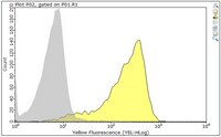
Quality AssuranceEvaluated by Flow Cytometry in C57BL/6 mouse splenocytes.

Flow Cytometry Analysis: 0.1 µg of this antibody detected H-2K immunoreactivity on the surface of one million C57BL/6 mouse splenocytes (H-2 haplotype b).
Usage Statement
  • Unless otherwise stated in our catalog or other company documentation accompanying the product(s), our products are intended for research use only and are not to be used for any other purpose, which includes but is not limited to, unauthorized commercial uses, in vitro diagnostic uses, ex vivo or in vivo therapeutic uses or any type of consumption or application to humans or animals.
Storage and Shipping Information
Storage ConditionsStable for 1 year at -20°C from date of receipt.
Handling Recommendations: Upon receipt and prior to removing the cap, centrifuge the vial and gently mix the solution. Aliquot into microcentrifuge tubes and store at -20°C. Avoid repeated freeze/thaw cycles, which may damage IgG and affect product performance.
Packaging Information
Material Size100 μg
Transport Information
Supplemental Information
Specifications
Global Trade Item Number
Catalogue Number GTIN
MABF946 04054839078767