Our broad portfolio consists of multiplex panels that allow you to choose, within the panel, analytes that best meet your needs. On a separate tab you can choose the premixed cytokine format or a single plex kit.
Cell Signaling Kits & MAPmates™
Choose fixed kits that allow you to explore entire pathways or processes. Or design your own kits by choosing single plex MAPmates™, following the provided guidelines.
The following MAPmates™ should not be plexed together:
-MAPmates™ that require a different assay buffer
-Phospho-specific and total MAPmate™ pairs, e.g. total GSK3β and GSK3β (Ser 9)
-PanTyr and site-specific MAPmates™, e.g. Phospho-EGF Receptor and phospho-STAT1 (Tyr701)
-More than 1 phospho-MAPmate™ for a single target (Akt, STAT3)
-GAPDH and β-Tubulin cannot be plexed with kits or MAPmates™ containing panTyr
.
Catalogue Number
Ordering Description
Qty/Pack
List
This item has been added to favorites.
Select A Species, Panel Type, Kit or Sample Type
To begin designing your MILLIPLEX® MAP kit select a species, a panel type or kit of interest.
Custom Premix Selecting "Custom Premix" option means that all of the beads you have chosen will be premixed in manufacturing before the kit is sent to you.
Catalogue Number
Ordering Description
Qty/Pack
List
This item has been added to favorites.
Species
Panel Type
Selected Kit
Qty
Catalogue Number
Ordering Description
Qty/Pack
List Price
96-Well Plate
Qty
Catalogue Number
Ordering Description
Qty/Pack
List Price
Add Additional Reagents (Buffer and Detection Kit is required for use with MAPmates)
Qty
Catalogue Number
Ordering Description
Qty/Pack
List Price
48-602MAG
Buffer Detection Kit for Magnetic Beads
1 Kit
Space Saver Option Customers purchasing multiple kits may choose to save storage space by eliminating the kit packaging and receiving their multiplex assay components in plastic bags for more compact storage.
This item has been added to favorites.
The Product Has Been Added To Your Cart
You can now customize another kit, choose a premixed kit, check out or close the ordering tool.
ST1202
Sigma-AldrichAnti-PGC-1α Mouse mAb (4C1.3)
This Anti-PGC-1α Mouse mAb (4C1.3) is validated for use in Immunoblotting, Immunocytochemistry, Immunoprecipitation, Paraffin Sections for the detection of PGC-1α.
More>>This Anti-PGC-1α Mouse mAb (4C1.3) is validated for use in Immunoblotting, Immunocytochemistry, Immunoprecipitation, Paraffin Sections for the detection of PGC-1α. Less<<
Anti-PGC-1α Mouse mAb (4C1.3) MSDS (material safety data sheet) or SDS, CoA and CoQ, dossiers, brochures and other available documents.
The quantity field is empty. Please enter a quantity of 1 or more to add items to your cart.
Description
Overview
Recognizes the endogenous forms of PGC-1α, which includes the ~113 kDa PGC-1α protein and the ~38 kDa splice variant, in brown adipose tissue, liver, and kidney.
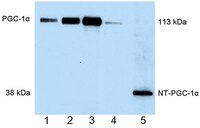
Detection of human PGC-1α by immunoblottiong. Samples: Whole cell lysates from COS cells transformed with wild-type PGC-1α (5 µg, lane 1; 10 µg, lane 2; 20 µg, lane 3), whole cell extract from brown adipose tissue of a cold-exposed mouse (lane 4), and whole cell lysate from CHO cells transformed with NT-PGC-1α (0.5 µg, lane 5). Primary antibody: Anti-PGC-1α Mouse mAb (4C1.3) (Cat. No. ST1202) (1 µg/ml). Detection chemiluminescence. Detection of mouse PGC-1α by immunoblottiong. Samples: NT-PGC-1α Positive Control, (lane 1), whole cell extract from brown adipose tissue of mice kept at room temperature (50 µg; lanes 2-4), whole cell extract from brown adipose tissue of mice exposed to 4°C for 5 h (50 µg; lanes 5-7), and PGC-1α positive control (lane 8). Primary antibody: Anti-PGC-1α Mouse mAb (4C1.3) (Cat. No. ST1202) (1 µg/ml). Detection: chemiluminescence. Detection of mouse PGC-1α by immunoblottiong. Samples: PGC-1α positive control (lane 1), nuclear extract from mouse liver (75 µg, lane 2), whole cell extract from mouse kidney (100 µg, lane 3), and whole cell extract from kidney of a PGC-1α knock-out mouse (100 µg, lane 4). Primary antibody: Anti-PGC-1α Mouse mAb (4C1.3) (Cat. No. ST1202) (1 µg/ml). Detection: chemiluminescence.
References
References
Lai, L., et al. 2008. Genes Dev. 14, 1948. Rodgers, J.T., et al. 2008. FEBS Lett. 582, 46. Mazzucotelli, A., et al. 2007. Diabetes 10, 2467. Nemoto, S., et al. 2005. J. Biol. Chem. 16, 16456.
Product Information
Form
Liquid
Formulation
One vial of 100 µg antibody in 50 mM PBS, see vial for lot-specific concentration. One vial of NT-PGC-1α Positive Control, supplied as 25 µl whole cell extract in RIPA buffer containing 10 mM &beta-mercaptoethanol and 1% SDS; load 10 µl per lane; add sample buffer prior to SDS-PAGE loading.
Endogenous PGC-1α is found primarily in the nucleus and NT-PGC-1α is found in both cytosolic and nuclear fractions. Antibody should be titrated for optimal results in individual systems.
Biological Information
Immunogen
a recombinant protein consisting of amino acids 1-120 of mouse PGC-1α
Immunogen
Mouse
Clone
4C1.3
Host
Mouse
Isotype
IgG
Species Reactivity
Human
Mouse
Rat
Antibody Type
Monoclonal Antibody
Safety Information
R Phrase
R: 21/22-36/37/38
Harmful in contact with skin and if swallowed. Irritating to eyes, respiratory system and skin.
S Phrase
S: 26-36/37-45
In case of contact with eyes, rinse immediately with plenty of water and seek medical advice. Wear suitable protective clothing and gloves. In case of accident or if you feel unwell, seek medical advice immediately (show the label where possible).
Storage and Shipping Information
Ship Code
Dry Ice Only
Toxicity
Multiple Toxicity Values, refer to MSDS
Storage
≤ -70°C
Avoid freeze/thaw
Avoid freeze/thaw
Do not freeze
Ok to freeze
Special Instructions
Following initial thaw, aliquot and freeze (-70°C).
Note that this data sheet is not lot-specific and is representative of the current specifications for this product. Please consult the vial label and the certificate of analysis for information on specific lots. Also note that shipping conditions may differ from storage conditions.
Detection of human PGC-1α by immunoblottiong. Samples: Whole cell lysates from COS cells transformed with wild-type PGC-1α (5 µg, lane 1; 10 µg, lane 2; 20 µg, lane 3), whole cell extract from brown adipose tissue of a cold-exposed mouse (lane 4), and whole cell lysate from CHO cells transformed with NT-PGC-1α (0.5 µg, lane 5). Primary antibody: Anti-PGC-1α Mouse mAb (4C1.3) (Cat. No. ST1202) (1 µg/ml). Detection chemiluminescence. Detection of mouse PGC-1α by immunoblottiong. Samples: NT-PGC-1α Positive Control, (lane 1), whole cell extract from brown adipose tissue of mice kept at room temperature (50 µg; lanes 2-4), whole cell extract from brown adipose tissue of mice exposed to 4°C for 5 h (50 µg; lanes 5-7), and PGC-1α positive control (lane 8). Primary antibody: Anti-PGC-1α Mouse mAb (4C1.3) (Cat. No. ST1202) (1 µg/ml). Detection: chemiluminescence. Detection of mouse PGC-1α by immunoblottiong. Samples: PGC-1α positive control (lane 1), nuclear extract from mouse liver (75 µg, lane 2), whole cell extract from mouse kidney (100 µg, lane 3), and whole cell extract from kidney of a PGC-1α knock-out mouse (100 µg, lane 4). Primary antibody: Anti-PGC-1α Mouse mAb (4C1.3) (Cat. No. ST1202) (1 µg/ml). Detection: chemiluminescence.
Description
Protein G purified mouse monclonal antibody. Recognizes the endogenous forms of PGC-1α, which includes the ~113 kDa PGC-1α protein and the ~38 kDa splice variant.
Background
PGC-1α is a co-activator of PPARα, PPARγ, and other transcription factors and regulates the transcriptonal program of adaptive thermogenesis in brown adipose tissue, hepatic/renal gluconeogenesis, and muscle fiber type switching. The full-length protein is 113 kDa and is induced in brown adipose tissue by cold exposure, in liver and kidney by fasting, and in skeletal muscle by exercise. Alternative splicing of the full-length gene produces a 270 aa N-terminal splice variant that migrates at ~38 kDa and is induced by the same physiological signals that induce expression of the full-length protein. The N-terminal PGC-1α splice variant is also subject to post translational modifications that alter its migration and apparent molecular weight.
Host
Mouse
Immunogen species
Mouse
Immunogen
a recombinant protein consisting of amino acids 1-120 of mouse PGC-1α
Clone
4C1.3
Isotype
IgG
Species
human, mouse, rat
Form
Liquid
Formulation
One vial of 100 µg antibody in 50 mM PBS, see vial for lot-specific concentration. One vial of NT-PGC-1α Positive Control, supplied as 25 µl whole cell extract in RIPA buffer containing 10 mM &beta-mercaptoethanol and 1% SDS; load 10 µl per lane; add sample buffer prior to SDS-PAGE loading.
Preservative
None
Comments
Endogenous PGC-1α is found primarily in the nucleus and NT-PGC-1α is found in both cytosolic and nuclear fractions. Antibody should be titrated for optimal results in individual systems.
Storage
Avoid freeze/thaw ≤ -70°C
Do Not Freeze
Ok to freeze
Special Instructions
Following initial thaw, aliquot and freeze (-70°C).
Toxicity
Multiple Toxicity Values, refer to MSDS
References
Lai, L., et al. 2008. Genes Dev. 14, 1948. Rodgers, J.T., et al. 2008. FEBS Lett. 582, 46. Mazzucotelli, A., et al. 2007. Diabetes 10, 2467. Nemoto, S., et al. 2005. J. Biol. Chem. 16, 16456.