Millipore Sigma Vibrant Logo
Attention: We have moved. Merck Millipore products are no longer available for purchase on MerckMillipore.com.Learn More

AP1063 PhosphoDetect™ Anti-PDH-E1α (pSer²³²) Rabbit pAb

AP1063
Purchase on Sigma-Aldrich

Overview

Replacement Information

Key Spec Table

Species ReactivityHostAntibody Type
H, M, RRbPolyclonal Antibody

Products

Catalogue NumberPackaging Qty/Pack
AP1063-50UG Plastová ampulka 50 μg
Description
Overview

This product has been discontinued.


Recognizes the ~44 kDa PDH-E1α protein phosphorylated at Ser232 in HEK293 cells.

Catalogue NumberAP1063
Brand Family Calbiochem®
SynonymsAnti-Pyruvate Dehydogenase pSer²³² Rabbit pAb
Application Data
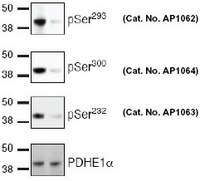
Detection of human PDH-E1α, phosphorylated at Ser293, Ser300, and Ser232 by immunoblotting. Samples: Whole tissue extract from mouse liver (20 µg) left untreated (lane 1) or treated with dichloroacetate (DCA, 5 mM for 4 h) (lane 2). Primary antibody: PhosphoDetect™ Anti PDH-E1α (pSer293) Rabbit pAb (Cat. No. AP1062) PhosphoDetect™ Anti PDH-E1α (pSer300) Rabbit pAb (Cat. No. AP1064) PhosphoDetect™ Anti PDH-E1α (pSer232) Rabbit pAb (Cat. No. AP1063) all used at 0.25 µg/ml and Anti-PDHE-1α. Detection: chemiluminescence. Data courtesy of Matthew Rardin, University of California San Diego.

Detection of PDH-E1α, phosphorylated at Ser293, Ser300, and Ser232 by immunocytochemistry. Samples: COS7 cells incubated in presence dichloroacetate (DCA; 5 mM for 2 h). All samples were incubated with MitoTracker® Red stain (100 nM for 20 min; to visualize mitochondria) and fixed in 3.7% formaldehyde, followed by methanol (3 min at -20°C), and permeabilization in PBS, 6% BSA, 0.3% Triton™ X-100 detergent, 0.1% Tween® 20 detergent. Primary antibodies: PhosphoDetect™ Anti-PDH-E1α (pSer293) Rabbit pAb (Cat. No. AP1062) (panel B), PhosphoDetect™ Anti-PDH-E1α (pSer300) Rabbit pAb (Cat. No. AP1064) (panel C), PhosphoDetect™ Anti-PDH-E1α (pSer232) Rabbit pAb (Cat. No. AP1063) (panel D) (0.5 µg/ml, green fluorescence) and anti-PDH-E1α antibody (green fluorescence, top 3 panels) (panel A). Detection: fluorescence (Alexa Fluor® 488 secondary antibody) with DAPI counterstain (300 nM, 1 min, blue fluorescence). Data courtesy of Matthew Rardin, University of California San Diego.
References
ReferencesRardin M.J., et. al. 2009. Anal. Biochem. 2, 157.
Seifert, F., et al. 2007. Biochemistry 21, 6277.
Lee, J., et al. 2007. Mol. Cell Prot. 4, 669.
Patel, M.S. and Korotchkina, L.G. 2006 Biochem. Soc. Trans. 34, 217.
Korotchkina, L.G., et al. 2001. J. Biol. Chem. 40, 37223.
Product Information
FormLiquid
FormulationIn PBS.
Positive controlHEK293 cells
Preservative≤0.1% sodium azide
Quality LevelMQ200
Applications
Key Applications Immunoblotting (Western Blotting)
Immunofluorescence
Immunoprecipitation
Application NotesImmunoblotting (0.25 µg/ml)
Immunofluorescence (Please see comments)
Immunoprecipitation (1 µg/ml)
Application CommentsThis antibody has been known to work with immunofluorescence. Antibody should be titrated for optimal results in individual systems.
Biological Information
Immunogena synthetic phosphopeptide corresponding to amino acids surrounding the Ser²³² phosphorylation site of human PDH-E1α
ImmunogenHuman
HostRabbit
Species Reactivity
  • Human
  • Mouse
  • Rat
Antibody TypePolyclonal Antibody
Physicochemical Information
Dimensions
Materials Information
Toxicological Information
Safety Information according to GHS
Safety Information
Product Usage Statements
Storage and Shipping Information
Ship Code Blue Ice Only
Toxicity Standard Handling
Storage -20°C
Avoid freeze/thaw Avoid freeze/thaw
Do not freeze Ok to freeze
Special InstructionsFollowing initial thaw, aliquot and freeze (-20°C).
Packaging Information
Transport Information
Supplemental Information
Specifications
Global Trade Item Number
Catalogue Number GTIN
AP1063-50UG 04055977227789

Documentation

PhosphoDetect™ Anti-PDH-E1α (pSer²³²) Rabbit pAb MSDS

Title

Safety Data Sheet (SDS) 

PhosphoDetect™ Anti-PDH-E1α (pSer²³²) Rabbit pAb Certificates of Analysis

TitleLot Number
AP1063

References

Reference overview
Rardin M.J., et. al. 2009. Anal. Biochem. 2, 157.
Seifert, F., et al. 2007. Biochemistry 21, 6277.
Lee, J., et al. 2007. Mol. Cell Prot. 4, 669.
Patel, M.S. and Korotchkina, L.G. 2006 Biochem. Soc. Trans. 34, 217.
Korotchkina, L.G., et al. 2001. J. Biol. Chem. 40, 37223.
Data Sheet

Note that this data sheet is not lot-specific and is representative of the current specifications for this product. Please consult the vial label and the certificate of analysis for information on specific lots. Also note that shipping conditions may differ from storage conditions.

Revision29-December-2010 RFH
SynonymsAnti-Pyruvate Dehydogenase pSer²³² Rabbit pAb
ApplicationImmunoblotting (0.25 µg/ml)
Immunofluorescence (Please see comments)
Immunoprecipitation (1 µg/ml)
Application Data
Detection of human PDH-E1α, phosphorylated at Ser293, Ser300, and Ser232 by immunoblotting. Samples: Whole tissue extract from mouse liver (20 µg) left untreated (lane 1) or treated with dichloroacetate (DCA, 5 mM for 4 h) (lane 2). Primary antibody: PhosphoDetect™ Anti PDH-E1α (pSer293) Rabbit pAb (Cat. No. AP1062) PhosphoDetect™ Anti PDH-E1α (pSer300) Rabbit pAb (Cat. No. AP1064) PhosphoDetect™ Anti PDH-E1α (pSer232) Rabbit pAb (Cat. No. AP1063) all used at 0.25 µg/ml and Anti-PDHE-1α. Detection: chemiluminescence. Data courtesy of Matthew Rardin, University of California San Diego.

Detection of PDH-E1α, phosphorylated at Ser293, Ser300, and Ser232 by immunocytochemistry. Samples: COS7 cells incubated in presence dichloroacetate (DCA; 5 mM for 2 h). All samples were incubated with MitoTracker® Red stain (100 nM for 20 min; to visualize mitochondria) and fixed in 3.7% formaldehyde, followed by methanol (3 min at -20°C), and permeabilization in PBS, 6% BSA, 0.3% Triton™ X-100 detergent, 0.1% Tween® 20 detergent. Primary antibodies: PhosphoDetect™ Anti-PDH-E1α (pSer293) Rabbit pAb (Cat. No. AP1062) (panel B), PhosphoDetect™ Anti-PDH-E1α (pSer300) Rabbit pAb (Cat. No. AP1064) (panel C), PhosphoDetect™ Anti-PDH-E1α (pSer232) Rabbit pAb (Cat. No. AP1063) (panel D) (0.5 µg/ml, green fluorescence) and anti-PDH-E1α antibody (green fluorescence, top 3 panels) (panel A). Detection: fluorescence (Alexa Fluor® 488 secondary antibody) with DAPI counterstain (300 nM, 1 min, blue fluorescence). Data courtesy of Matthew Rardin, University of California San Diego.
DescriptionImmunoaffinity purified rabbit polyclonal antibody. Recognizes the ~44 kDa PDH-E1α protein phosphorylated at Ser232.
BackgroundThe pyruvate dehydogenase (PDH) complex (PDC) catalyzes the irreversible conversion of pyruvate to acetyl CoA, providing the link between glycolyis and the TCA cycle. Dysregulation of the PDC in skeletal muscle has been implicated in type 2 diabetes and mutations in the PDC result in various mitochondrial diseases. The PDC is a multienzyme complex composed of several copies of three catalytic proteins (E1, E2, and E3): one structural (E3-binding protein) and two regulatory proteins (PDH kinase [PDK] and PDH phosphatase [PDP]). The activity of the PDC is mainly regulated by phosphorylation-dephosphorylation of three specific serine residues (232, 293, 300) on the E1 subunit. Phosphorylation of PDH determines the activity of the complex. Phosphorylation by one of four PDK isoforms inactivates the complex, while removal of the phosphate by one of two PDP isoforms activates the complex. The activity of the PDP1 isoform is sensitive to Ca2+, while the PDK isoforms are regulated by the reduction and acetylation status of the E2 subunit, pyruvate, and the small molecule the anti cancer dichloroacetate. (DCA).
HostRabbit
Immunogen speciesHuman
Immunogena synthetic phosphopeptide corresponding to amino acids surrounding the Ser²³² phosphorylation site of human PDH-E1α
Specieshuman, mouse, rat
Positive controlHEK293 cells
FormLiquid
FormulationIn PBS.
Preservative≤0.1% sodium azide
CommentsThis antibody has been known to work with immunofluorescence. Antibody should be titrated for optimal results in individual systems.
Storage Avoid freeze/thaw
-20°C
Do Not Freeze Ok to freeze
Special InstructionsFollowing initial thaw, aliquot and freeze (-20°C).
Toxicity Standard Handling
ReferencesRardin M.J., et. al. 2009. Anal. Biochem. 2, 157.
Seifert, F., et al. 2007. Biochemistry 21, 6277.
Lee, J., et al. 2007. Mol. Cell Prot. 4, 669.
Patel, M.S. and Korotchkina, L.G. 2006 Biochem. Soc. Trans. 34, 217.
Korotchkina, L.G., et al. 2001. J. Biol. Chem. 40, 37223.