Trypanosoma cruzi coaxes cardiac fibroblasts into preventing cardiomyocyte death by activating nerve growth factor receptor TrkA.
Aridgides, D; Salvador, R; PereiraPerrin, M
PloS one
8
e57450
2013
Show Abstract
Cardiomyocytes express neurotrophin receptor TrkA that promotes survival following nerve growth factor (NGF) ligation. Whether TrkA also resides in cardiac fibroblasts (CFs) and underlies cardioprotection is unknown.To test whether CFs express TrkA that conveys paracrine signals to neighbor cardiomyocytes using, as probe, the Chagas disease parasite Trypanosoma cruzi, which expresses a TrkA-binding neurotrophin mimetic, named PDNF. T. cruzi targets the heart, causing chronic debilitating cardiomyopathy in ∼30% patients.Basal levels of TrkA and TrkC in primary CFs are comparable to those in cardiomyocytes. However, in the myocardium, TrkA expression is significantly lower in fibroblasts than myocytes, and vice versa for TrkC. Yet T. cruzi recognition of TrkA on fibroblasts, preferentially over cardiomyocytes, triggers a sharp and sustained increase in NGF, including in the heart of infected mice or of mice administered PDNF intravenously, as early as 3-h post-administration. Further, NGF-containing T. cruzi- or PDNF-induced fibroblast-conditioned medium averts cardiomyocyte damage by H(2)O(2), in agreement with the previously recognized cardioprotective role of NGF.TrkA residing in CFs induces an exuberant NGF production in response to T. cruzi infection, enabling, in a paracrine fashion, myocytes to resist oxidative stress, a leading Chagas cardiomyopathy trigger. Thus, PDNF-TrkA interaction on CFs may be a mechanism orchestrated by T. cruzi to protect its heart habitat, in concert with the long-term (decades) asymptomatic heart parasitism that characterizes Chagas disease. Moreover, as a potent booster of cardioprotective NGF in vivo, PDNF may offer a novel therapeutic opportunity against cardiomyopathies. | 23437390
 |
Artemin causes hypersensitivity to warm sensation, mimicking warmth-provoked pruritus in atopic dermatitis.
Hiroyuki Murota,Mayuko Izumi,Mostafa I A Abd El-Latif,Megumi Nishioka,Mika Terao,Mamori Tani,Saki Matsui,Shigetoshi Sano,Ichiro Katayama
The Journal of allergy and clinical immunology
130
2011
Show Abstract
Itch impairs the quality of life for many patients with dermatoses, especially atopic dermatitis (AD), and is frequently induced by a warm environment. | 22770266
 |
Development of N-methyl-D-aspartate receptor subunit immunoreactivity in the neonatal gerbil cochlear nucleus.
D Joelson, I R Schwartz
Microscopy research and technique
41
246-62
1998
Show Abstract
The distribution of immunoreactivity for the ionotropic N-methyl-D-aspartate (NMDA) receptor subunits was mapped in the cochlear nucleus of postnatal day (P) 7, P14, P21, and P28 gerbils. Frozen sections and serial plastic sections of tissue were incubated with antibodies to NMDAR1 (NR1), NMDAR2A (NR2A), NMDAR2A/B (NR2A/B), and NMDAR2B (NR2B). An overall diffuse stain was noted at P7 for NR1 and NR2A/B. Staining of neuronal somata in the dorsal cochlear nucleus molecular layer and fusiform cell layer, the posteroventral cochlear nucleus octopus cell area, and the anteroventral cochlear nucleus increased from P7 to P28. Staining of the neuropil (the unresolved mass of processes and axons, excluding only neuronal somata and distinctly stained proximal dendrites) of the deep dorsal cochlear nucleus and posteroventral cochlear nucleus showed a steady decrease, while molecular layer neuropil remained moderately stained. The NR2A antibody produced a distinctive staining of dendrites in the dorsal cochlear nucleus deep and fusiform cell layers seen first at P14 with increasing dendritic lengths stained at P21 and P28. Giant neurons of the deep dorsal cochlear nucleus were the most conspicuous somata stained by the NR2A. Their stained dendrites spanned much of the dorsal cochlear nucleus deep and fusiform cell layers and even extended into the octopus cell area of the posteroventral cochlear nucleus. Dendritic staining was also present in caudal and rostral posteroventral cochlear nucleus, first distinguishable at P14 and becoming increasingly strong. The Chemicon polyclonal NR2B antibody produced glial staining especially prominent in the caudal posteroventral cochlear nucleus and the dorsal cochlear nucleus fusiform cell layer, most intense at P7 and subsequently decreasing, although not disappearing, in all areas through P28. The Molecular Probes (Eugene, OR) polyclonal NR2B produced a light granular staining pattern over a number of somata but no glial staining. Neuropil staining was not prominent with either NR2B antibody. Differences in changes of neonatal immunoreactivity patterns in different populations of cochlear nucleus neuronal somata and dendrites for NR1, NR2A, NR2A/B, and NR2B suggest that alterations in some receptor composition is occurring over the period spanning the onset of hearing. | 9605342
 |
Denervation, but not decentralization, reduces nerve growth factor content of the mesenteric artery.
Liu, D T, et al.
J. Neurochem., 66: 2295-9 (1996)
1996
Show Abstract
In the present study we applied an improved nerve growth factor (NGF) extraction method to examine the effects of denervation and sympathetic decentralization on NGF levels in vascular tissue. Adult male Wistar Kyoto rats underwent mesenteric arterial denervation or splanchnic nerve transection. Four days after operation, animals were killed, and the mesenteric artery and coeliac-superior mesenteric ganglia were removed. The arterial adventitia was stripped from the media to measure NGF levels in nerve and smooth muscle separately. A high concentration of NGF was detected in the normal artery, 90% of which was in the adventitial layer. Surgical denervation significantly reduced the NGF levels in the artery and ganglia by 78 and 71%, respectively. However, within the artery the level of NGF was reduced in the adventitia but not in the media. Thus, the large reduction of NGF content resulted from the loss of nerve plexus from the artery. In contrast, decentralization did not alter the NGF content in the artery, in either the adventitia or media. Our results are in marked contrast to previous studies reporting elevated levels of NGF following denervation. This discrepancy is explained by the ability of our new procedure to extract much greater amounts of NGF from the tissue. | 8632151
 |
An improved procedure for the immunohistochemical localization of nerve growth factor-like immunoreactivity.
Zhou, X F, et al.
J. Neurosci. Methods, 54: 95-102 (1994)
1993
Show Abstract
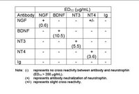
Nerve growth factor (NGF) is a survival factor required by a number of neuronal populations including most post-ganglionic sympathetic neurones. NGF has been detected and quantified in many tissues but there is little information regarding its cellular localization. Although it has been argued that histological detection has proven difficult due to the low levels of NGF present, other factors may contribute to prevent its identification. In the present study, we report a method for the histological detection of NGF-like immunoreactivity in the rat superior cervical ganglia (SCG). Adult Wistar-Kyoto rats were perfused briefly with either a high or low pH buffer prior to fixation and routine immunohistochemistry. Polyclonal antibodies to native mouse NGF used in the present study recognized mouse NGF but not recombinant human neurotrophin 3 (rhNT3) or brain-derived neurotrophic factor (rhBDNF) by immunoblot analysis. NGF-like immunoreactivity was localized to most sympathetic neurones. Immunoreactivity was detected in the cytoplasm with dense labelling around nuclei. No stain was seen in sections incubated with normal sheep IgG or from animals perfused with phosphate buffer (pH 7.4) prior to fixation. In addition, axotomy resulted in the disappearance of NGF immunoreactivity which was confirmed by biochemical quantification. Finally, no NGF immunoreactivity was found in neurones of rats treated systemically with NGF antiserum 3 days earlier. Possible mechanisms underlying the improvement of NGF immunohistochemistry by pH manipulation before fixation are discussed. | 7815824
 |
Transport of endogenous nerve growth factor in the proximal stump of sectioned nerves.
Abrahamson, I K, et al.
J. Neurocytol., 16: 417-22 (1987)
1987
Show Abstract
Immunohistochemistry has been used to demonstrate the presence of nerve growth factor (NGF)-like immunoreactivity in normal and sectioned mouse sciatic nerves. In normal nerves, immunoreactive material was not visible unless a silk ligature had previously been applied to constrict the nerves, and only then in the segment of nerve immediately distal to the ligation. Immunoreactivity was visible as early as 2 h after application of the ligature. When nerves were sectioned prior to ligation to prevent the transport of material from nerve terminals within innervated tissues, the NGF-like immunoreactivity continued to accumulate. This accumulation also occurred when a portion of the proximal stump from sectioned nerves was removed from the animal and placed in culture. Quantitative estimate of NGF concentrations with a sensitive immunoassay showed that the amount of NGF present within a segment of the proximal stump of sectioned nerves more than doubled in a 24 h period. The findings indicate that NGF is produced by cells within sectioned nerves, and further suggest that in the normal intact nerve at least a proportion of the NGF being transported derives from these cells. | 2441000
 |


