Millipore Sigma Vibrant Logo
Attention: We have moved. Merck Millipore products are no longer available for purchase on MerckMillipore.com.Learn More

NE1018 Anti-Myelin Basic Protein Mouse mAb (SMI-94)

NE1018
  
Purchase on Sigma-Aldrich

Overview

Replacement Information

Key Spec Table

Species ReactivityHostAntibody Type
Ca, H, Mk, M, Rb, RMMonoclonal Antibody
Description
Overview

This product has been discontinued.


Recognizes the ~21 kDa myelin basic protein in rat brain tissue.

Catalogue NumberNE1018
Brand Family Calbiochem®
Application Data
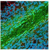
Detection of rat myelin basic protein by staining frozen sections. Sample: Rat brain. Primary antibody: Anti-Myelin Basic Protein Mouse mAb (SMI-94) (Cat. No. NE1018) (1:1000). Detection: fluorescence (green) with Hoechst counterstain.
References
ReferencesEvers, P. and Uylings, H.B. 1997. J. Neurosci. Methods 72, 197.
Shin, R.W., et al. 1991. Lab. Invest. 64, 693.
Product Information
FormLiquid
FormulationUndiluted ascites.
Positive controlRat brain
Preservative≤0.1% sodium azide
Applications
Key Applications Enzyme-Linked Immunosorbent Assay
Frozen Sections
Immunoblotting (Western Blotting)
Immunocytochemistry
Paraffin Sections
Application NotesELISA (1:1000)
Frozen Sections (1:1000, see comments)
Immunoblotting (1:1000)
Immunocytochemistry (1:1000)
Paraffin Sections (1:1000, heat pre-treatment required, see comments)
Application CommentsOther species have not been tested, but the antibody is expected to recognize MBP in most mammalian species. Recognizes developing and adult myelin and developing oligodendrocytes. Distinguishes oligodendrocytes from astrocytes, microglia, neurons, and other cells in brain tissue. In conjunction with Anti-Myelin Basic Protein Mouse mAb (SMI-91) (Cat. No. NE1020) and/or Anti-Myelin Basic Protein Mouse mAb (SMI-99) (Cat. No. NE1019), this antibody is useful for immunocytochemical studies on the progression of normal and pathologic myelination. Tissues can be fixed in a variety of paraformaldehyde- or formaldehyde-containing fixatives. To insure penetration of the antibody into myelin it is necessary to pre-treat frozen sections with cold methanol for 30 min or detergent. For staining paraffin sections it is recommended that de-paraffinized tissues be autoclaved in dH2O for 10 min or boiled in Tris-saline, pH 9.0, in a microwave for 15 min to expose the epitope. Antibody should be titrated for optimal results in individual systems.
Biological Information
Immunogenpurified human myelin basic protein
ImmunogenHuman
EpitopeWithin amino acids 70-89
CloneSMI-94
HostMouse
IsotypeIgG₁
Species Reactivity
  • Canine
  • Human
  • Monkey
  • Mouse
  • Rabbit
  • Rat
Antibody TypeMonoclonal Antibody
Physicochemical Information
Dimensions
Materials Information
Toxicological Information
Safety Information according to GHS
Safety Information
Product Usage Statements
Storage and Shipping Information
Ship Code Dry Ice Only
Toxicity Standard Handling
Storage -20°C
Avoid freeze/thaw Avoid freeze/thaw
Do not freeze Ok to freeze
Special InstructionsFollowing initial thaw, aliquot and freeze (-20°C).
Packaging Information
Transport Information
Supplemental Information
Specifications
Global Trade Item Number
Catalogue Number GTIN
NE1018 0

Documentation

Anti-Myelin Basic Protein Mouse mAb (SMI-94) MSDS

Title

Safety Data Sheet (SDS) 

Anti-Myelin Basic Protein Mouse mAb (SMI-94) Certificates of Analysis

TitleLot Number
NE1018

References

Reference overview
Evers, P. and Uylings, H.B. 1997. J. Neurosci. Methods 72, 197.
Shin, R.W., et al. 1991. Lab. Invest. 64, 693.
Data Sheet

Note that this data sheet is not lot-specific and is representative of the current specifications for this product. Please consult the vial label and the certificate of analysis for information on specific lots. Also note that shipping conditions may differ from storage conditions.

Revision01-October-2007 RFH
ApplicationELISA (1:1000)
Frozen Sections (1:1000, see comments)
Immunoblotting (1:1000)
Immunocytochemistry (1:1000)
Paraffin Sections (1:1000, heat pre-treatment required, see comments)
Application Data
Detection of rat myelin basic protein by staining frozen sections. Sample: Rat brain. Primary antibody: Anti-Myelin Basic Protein Mouse mAb (SMI-94) (Cat. No. NE1018) (1:1000). Detection: fluorescence (green) with Hoechst counterstain.
DescriptionMouse monoclonal antibody supplied as undiluted ascites. Recognizes the ~21 kDa myelin basic protein (MBP).
BackgroundThe myelin sheath is essential for rapid and effective propagation of action potential within the axon. Mutations identified in several myelin-related genes reportedly lead to severe demyelinating neuropathies. Recent evidence also suggests that the autoimmune disease Multiple Sclerosis (MS) is mediated by T cells with specificity for antigens in the myelin sheath. Although myelin in the central nervous system (CNS) and peripheral nervous system (PNS) is structurally similar, it is chemically distinct since oligodendrocytes form myelin in the CNS whereas Schwann cells generate PNS myelin. Myelin proteolipid protein (PLP) is the predominant integral membrane protein in mammalian CNS myelin. In contrast, myelin basic protein (MBP) is localized to the myelin cytoplasm and is highly conserved among species. Both MBP and PLP mediate myelin compaction, whereas thickness is regulated by MBP. Several other myelin proteins have been identified to play distinct or partially overlapping functions in the formation and maintenance of the myelin sheath, including P0, myelin-associated glycoprotein (MAG), peripheral myelin protein (PMP22), and Connexin 32 (Cx32). The development of the myelin sheath has been a good model for understanding the mechanisms of neuron-glia interaction.
HostMouse
Immunogen speciesHuman
Immunogenpurified human myelin basic protein
EpitopeWithin amino acids 70-89
CloneSMI-94
IsotypeIgG₁
Speciescanine, human, monkey, mouse, rabbit, rat
Positive controlRat brain
FormLiquid
FormulationUndiluted ascites.
Preservative≤0.1% sodium azide
CommentsOther species have not been tested, but the antibody is expected to recognize MBP in most mammalian species. Recognizes developing and adult myelin and developing oligodendrocytes. Distinguishes oligodendrocytes from astrocytes, microglia, neurons, and other cells in brain tissue. In conjunction with Anti-Myelin Basic Protein Mouse mAb (SMI-91) (Cat. No. NE1020) and/or Anti-Myelin Basic Protein Mouse mAb (SMI-99) (Cat. No. NE1019), this antibody is useful for immunocytochemical studies on the progression of normal and pathologic myelination. Tissues can be fixed in a variety of paraformaldehyde- or formaldehyde-containing fixatives. To insure penetration of the antibody into myelin it is necessary to pre-treat frozen sections with cold methanol for 30 min or detergent. For staining paraffin sections it is recommended that de-paraffinized tissues be autoclaved in dH2O for 10 min or boiled in Tris-saline, pH 9.0, in a microwave for 15 min to expose the epitope. Antibody should be titrated for optimal results in individual systems.
Storage Avoid freeze/thaw
-20°C
Do Not Freeze Ok to freeze
Special InstructionsFollowing initial thaw, aliquot and freeze (-20°C).
Toxicity Standard Handling
ReferencesEvers, P. and Uylings, H.B. 1997. J. Neurosci. Methods 72, 197.
Shin, R.W., et al. 1991. Lab. Invest. 64, 693.