Targeting the LOX/hypoxia axis reverses many of the features that make pancreatic cancer deadly: inhibition of LOX abrogates metastasis and enhances drug efficacy.
Miller, BW; Morton, JP; Pinese, M; Saturno, G; Jamieson, NB; McGhee, E; Timpson, P; Leach, J; McGarry, L; Shanks, E; Bailey, P; Chang, D; Oien, K; Karim, S; Au, A; Steele, C; Carter, CR; McKay, C; Anderson, K; Evans, TR; Marais, R; Springer, C; Biankin, A; Erler, JT; Sansom, OJ
EMBO molecular medicine
7
1063-76
2015
Show Abstract
Pancreatic ductal adenocarcinoma (PDAC) is one of the leading causes of cancer-related mortality. Despite significant advances made in the treatment of other cancers, current chemotherapies offer little survival benefit in this disease. Pancreaticoduodenectomy offers patients the possibility of a cure, but most will die of recurrent or metastatic disease. Hence, preventing metastatic disease in these patients would be of significant benefit. Using principal component analysis (PCA), we identified a LOX/hypoxia signature associated with poor patient survival in resectable patients. We found that LOX expression is upregulated in metastatic tumors from Pdx1-Cre Kras(G12D/+) Trp53(R172H/+) (KPC) mice and that inhibition of LOX in these mice suppressed metastasis. Mechanistically, LOX inhibition suppressed both migration and invasion of KPC cells. LOX inhibition also synergized with gemcitabine to kill tumors and significantly prolonged tumor-free survival in KPC mice with early-stage tumors. This was associated with stromal alterations, including increased vasculature and decreased fibrillar collagen, and increased infiltration of macrophages and neutrophils into tumors. Therefore, LOX inhibition is able to reverse many of the features that make PDAC inherently refractory to conventional therapies and targeting LOX could improve outcome in surgically resectable disease. | 26077591
 |
Attenuation of lysyl oxidase and collagen gene expression in keratoconus patient corneal epithelium corresponds to disease severity.
Shetty, R; Sathyanarayanamoorthy, A; Ramachandra, RA; Arora, V; Ghosh, A; Srivatsa, PR; Pahuja, N; Nuijts, RM; Sinha-Roy, A; Mohan, RR; Ghosh, A
Molecular vision
21
12-25
2015
Show Abstract
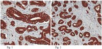
Keratoconus (KC) is characterized by progressive vision loss due to corneal thinning and structural abnormalities. It is hypothesized that KC is caused by deregulated collagen levels and collagen fibril-maturating enzyme lysyl oxidase (LOX). Further, it is currently not understood whether the gene expression deregulated by the corneal epithelium influences KC pathogenesis. We studied (i) the expressions of the LOX, collagen I (COL IA1), collagen IV (COL IVA1), MMP9, and IL6 genes in KC corneal epithelia, (ii) validated their expression levels in patient tissues, and (iii) correlated expression levels with KC disease severity. The primary goal of this study was to evaluate the importance of these genes in the progression of KC.We analyzed the gene expression levels of the key proteins LOX, collagens (COL IA1 and COL IVA1), MMP9, and IL6 in debrided corneal epithelia from a large cohort of KC patients (90 eyes) and compared them to control patients (52 eyes) without KC. We measured the total LOX activity in the tears of KC patients compared to controls. We also correlated the protein expression levels of LOX and collagens by immunohistochemistry (IHC) in primary tissues from KC patients (27 eyes) undergoing keratoplasty compared to healthy donor corneas (15 eyes).We observed a significant reduction in LOX transcript levels in KC corneal epithelia, and LOX activity in KC tears correlated with disease severity. Collagen transcripts were also reduced in KC while MMP9 transcript levels were upregulated and correlated with disease severity. IL6 was moderately increased in KC patients. IHC demonstrated a reduction in the protein expression levels of LOX in the epithelium and collagen IV in the basement membrane of KC patients compared to healthy donor corneas.The data demonstrates that the structural deformity of the KC cornea may be dependent on reduced expressions of collagens and LOX, as well as on MMP9 elevated by the corneal epithelium. | 25593510
|