Saccadic Palsy following Cardiac Surgery: Possible Role of Perineuronal Nets.
Eggers, SD; Horn, AK; Roeber, S; Härtig, W; Nair, G; Reich, DS; Leigh, RJ
PloS one
10
e0132075
2015
Show Abstract
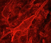
Perineuronal nets (PN) form a specialized extracellular matrix around certain highly active neurons within the central nervous system and may help to stabilize synaptic contacts, promote local ion homeostasis, or play a protective role. Within the ocular motor system, excitatory burst neurons and omnipause neurons are highly active cells that generate rapid eye movements - saccades; both groups of neurons contain the calcium-binding protein parvalbumin and are ensheathed by PN. Experimental lesions of excitatory burst neurons and omnipause neurons cause slowing or complete loss of saccades. Selective palsy of saccades in humans is reported following cardiac surgery, but such cases have shown normal brainstem neuroimaging, with only one clinicopathological study that demonstrated paramedian pontine infarction. Our objective was to test the hypothesis that lesions of PN surrounding these brainstem saccade-related neurons may cause saccadic palsy.Together with four controls we studied the brain of a patient who had developed a permanent selective saccadic palsy following cardiac surgery and died several years later. Sections of formalin-fixed paraffin-embedded brainstem blocks were applied to double-immunoperoxidase staining of parvalbumin and three different components of PN. Triple immunofluorescence labeling for all PN components served as internal controls. Combined immunostaining of parvalbumin and synaptophysin revealed the presence of synapses.Excitatory burst neurons and omnipause neurons were preserved and still received synaptic input, but their surrounding PN showed severe loss or fragmentation.Our findings support current models and experimental studies of the brainstem saccade-generating neurons and indicate that damage to PN may permanently impair the function of these neurons that the PN ensheathe. How a postulated hypoxic mechanism could selectively damage the PN remains unclear. We propose that the well-studied saccadic eye movement system provides an accessible model to evaluate the role of PN in health and disease. | | 26135580
 |
Restricted loss of olivocochlear but not vestibular efferent neurons in the senescent gerbil (Meriones unguiculatus).
Radtke-Schuller, S; Seeler, S; Grothe, B
Frontiers in aging neuroscience
7
4
2015
Show Abstract
Degeneration of hearing and vertigo are symptoms of age-related auditory and vestibular disorders reflecting multifactorial changes in the peripheral and central nervous system whose interplay remains largely unknown. Originating bilaterally in the brain stem, vestibular and auditory efferent cholinergic projections exert feedback control on the peripheral sensory organs, and modulate sensory processing. We studied age-related changes in the auditory and vestibular efferent systems by evaluating number of cholinergic efferent neurons in young adult and aged gerbils, and in cholinergic trigeminal neurons serving as a control for efferents not related to the inner ear. We observed a significant loss of olivocochlear (OC) neurons in aged compared to young adult animals, whereas the overall number of lateral superior olive (LSO) cells was not reduced in aging. Although the loss of lateral and medial olivocochlear (MOC) neurons was uniform and equal on both sides of the brain, there were frequency-related differences within the lateral olivocochlear (LOC) neurons, where the decline was larger in the medial limb of the superior olivary nucleus (high frequency representation) than in the lateral limb (middle-to-low frequency representation). In contrast, neither the number of vestibular efferent neurons, nor the population of motor trigeminal neurons were significantly reduced in the aged animals. These observations suggest differential effects of aging on the respective cholinergic efferent brainstem systems. | | 25762929
 |
Aggrecan and chondroitin-6-sulfate abnormalities in schizophrenia and bipolar disorder: a postmortem study on the amygdala.
Pantazopoulos, H; Markota, M; Jaquet, F; Ghosh, D; Wallin, A; Santos, A; Caterson, B; Berretta, S
Translational psychiatry
5
e496
2015
Show Abstract
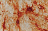
Perineuronal nets (PNNs) are specialized extracellular matrix aggregates surrounding distinct neuronal populations and regulating synaptic functions and plasticity. Previous findings showed robust PNN decreases in amygdala, entorhinal cortex and prefrontal cortex of subjects with schizophrenia (SZ), but not bipolar disorder (BD). These studies were carried out using a chondroitin sulfate proteoglycan (CSPG) lectin marker. Here, we tested the hypothesis that the CSPG aggrecan, and 6-sulfated chondroitin sulfate (CS-6) chains highly represented in aggrecan, may contribute to these abnormalities. Antibodies against aggrecan and CS-6 (3B3 and CS56) were used in the amygdala of healthy control, SZ and BD subjects. In controls, aggrecan immunoreactivity (IR) was observed in PNNs and glial cells. Antibody 3B3, but not CS56, also labeled PNNs in the amygdala. In addition, dense clusters of CS56 and 3B3 IR encompassed CS56- and 3B3-IR glia, respectively. In SZ, numbers of aggrecan- and 3B3-IR PNNs were decreased, together with marked reductions of aggrecan-IR glial cells and CS-6 (3B3 and CS56)-IR 'clusters'. In BD, numbers of 3B3-IR PNNs and CS56-IR clusters were reduced. Our findings show disruption of multiple PNN populations in the amygdala of SZ and, more modestly, BD. Decreases of aggrecan-IR glia and CS-6-IR glial 'clusters', in sharp contrast to increases of CSPG/lectin-positive glia previously observed, indicate that CSPG abnormalities may affect distinct glial cell populations and suggest a potential mechanism for PNN decreases. Together, these abnormalities may contribute to a destabilization of synaptic connectivity and regulation of neuronal functions in the amygdala of subjects with major psychoses. | | 25603412
 |
Chondroitin sulfate proteoglycans potently inhibit invasion and serve as a central organizer of the brain tumor microenvironment.
Silver, DJ; Siebzehnrubl, FA; Schildts, MJ; Yachnis, AT; Smith, GM; Smith, AA; Scheffler, B; Reynolds, BA; Silver, J; Steindler, DA
The Journal of neuroscience : the official journal of the Society for Neuroscience
33
15603-17
2013
Show Abstract
Glioblastoma (GBM) remains the most pervasive and lethal of all brain malignancies. One factor that contributes to this poor prognosis is the highly invasive character of the tumor. GBM is characterized by microscopic infiltration of tumor cells throughout the brain, whereas non-neural metastases, as well as select lower grade gliomas, develop as self-contained and clearly delineated lesions. Illustrated by rodent xenograft tumor models as well as pathological human patient specimens, we present evidence that one fundamental switch between these two distinct pathologies--invasion and noninvasion--is mediated through the tumor extracellular matrix. Specifically, noninvasive lesions are associated with a rich matrix containing substantial amounts of glycosylated chondroitin sulfate proteoglycans (CSPGs), whereas glycosylated CSPGs are essentially absent from diffusely infiltrating tumors. CSPGs, acting as central organizers of the tumor microenvironment, dramatically influence resident reactive astrocytes, inducing their exodus from the tumor mass and the resultant encapsulation of noninvasive lesions. Additionally, CSPGs induce activation of tumor-associated microglia. We demonstrate that the astrogliotic capsule can directly inhibit tumor invasion, and its absence from GBM presents an environment favorable to diffuse infiltration. We also identify the leukocyte common antigen-related phosphatase receptor (PTPRF) as a putative intermediary between extracellular glycosylated CSPGs and noninvasive tumor cells. In all, we present CSPGs as critical regulators of brain tumor histopathology and help to clarify the role of the tumor microenvironment in brain tumor invasion. | Immunohistochemistry | 24068827
 |
Alterations in chondroitin sulfate proteoglycan expression occur both at and far from the site of spinal contusion injury.
Andrews, EM; Richards, RJ; Yin, FQ; Viapiano, MS; Jakeman, LB
Experimental neurology
235
174-87
2011
Show Abstract
Chondroitin sulfate proteoglycans (CSPGs) present an inhibitory barrier to axonal growth and plasticity after trauma to the central nervous system. These extracellular and membrane bound molecules are altered after spinal cord injuries, but the magnitude, time course, and patterns of expression following contusion injury have not been fully described. Western blots and immunohistochemistry were combined to assess the expression of four classically inhibitory CSPGs, aggrecan, neurocan, brevican and NG2, at the lesion site and in distal segments of cervical and thoracic spinal cord at 3, 7, 14 and 28 days following a severe mid-thoracic spinal contusion. Total neurocan and the full-length (250 kDa) isoform were strongly upregulated both at the lesion epicenter and in cervical and lumbar segments. In contrast, aggrecan and brevican were sharply reduced at the injury site and were unchanged in distal segments. Total NG2 protein was unchanged across the injury site, while NG2+ profiles were distributed throughout the lesion site by 14 days post-injury (dpi). Far from the lesion, NG2 expression was increased at lumbar, but not cervical spinal cord levels. To determine if the robust increase in neurocan at the distal spinal cord levels corresponded to regions of increased astrogliosis, neurocan and GFAP immunoreactivity were measured in gray and white matter regions of the spinal enlargements. GFAP antibodies revealed a transient increase in reactive astrocyte staining in cervical and lumbar cord, peaking at 14 dpi. In contrast, neurocan immunoreactivity was specifically elevated in the cervical dorsal columns and in the lumbar ventral horn and remained high through 28 dpi. The long lasting increase of neurocan in gray matter regions at distal levels of the spinal cord may contribute to the restriction of plasticity in the chronic phase after SCI. Thus, therapies targeted at altering this CSPG both at and far from the lesion site may represent a reasonable addition to combined strategies to improve recovery after SCI. | | 21952042
 |
Perisynaptic aggrecan-based extracellular matrix coats in the human lateral geniculate body devoid of perineuronal nets.
D Lendvai,M Morawski,G Brückner,L Négyessy,G Baksa,T Glasz,L Patonay,R T Matthews,T Arendt,A Alpár
Journal of neuroscience research
90
2011
Show Abstract
The extracellular matrix surrounds different neuronal compartments in the mature nervous system. In a variety of vertebrates, most brain regions are loaded with a distinct type of extracellular matrix around the somatodendritic part of neurons, termed perineuronal nets. The present study reports that chondrotin sulfate proteoglycan-based matrix is structured differently in the human lateral geniculate body. Using various chondrotin sulfate proteoglycan-based extracellular matrix antibodies, we show that perisomatic matrix labeling is rather weak or absent, whereas dendrites are contacted by axonal coats appearing as small, oval structures. Confocal laser scanning microscopy and electron microscopy demonstrated that these typical structures are associated with synaptic loci on dendrites. Using multiple labelings, we show that different chondrotin sulfate proteoglycan components of the extracellular matrix do not associate exclusively with neuronal structures but possibly associate with glial structures as well. Finally, we confirm and extend previous findings in primates that intensity differences of various extracellular matrix markers between magno- and parvocellular layers reflect functional segregation between these layers in the human lateral geniculate body. | | 21959900
 |
Molecular compartmentalization of lateral geniculate nucleus in the gray squirrel (Sciurus carolinensis).
Felch, DL; Van Hooser, SD
Frontiers in neuroanatomy
6
12
2011
Show Abstract
Previous research has suggested that the three physiologically defined relay cell-types in mammalian lateral geniculate nucleus (LGN)-called parvocellular (P), magnocellular (M), and koniocellular (K) cells in primates and X, Y, and W cells in other mammals-each express a unique combination of cell-type marker proteins. However, some of the relationships among physiological classification and protein expression found in primates, prosimians, and tree shrews do not apply to carnivores and murid rodents. It remains unknown whether these are exceptions to a common rule for all mammals, or whether these relationships vary over a wide range of species. To address this question, we examined protein expression in the gray squirrel (Sciurus carolinensis), a highly visual rodent. Unlike many rodents, squirrel LGN is well laminated, and the organization of X-like, Y-like, and W-like cells relative to the LGN layers has been characterized physiologically. We labeled tissue sections through visual thalamus with antibodies to calbindin and parvalbumin, the antibody Cat-301, and the lectin WFA. Calbindin expression was found in W-like cells in LGN layer 3, just adjacent to the optic tract. These results suggest that calbindin is a common marker for the konicellular pathway in mammals. However, while parvalbumin expression characterizes P and M cells in primates and X and Y cells in tree shrews, here it identifies only about half of the X-like cells in LGN layers 1 and 2. Putative Y/M cell markers did not differentiate relay cells in this animal. Together, these results suggest that protein expression patterns among LGN relay cell classes are variable across mammals. | | 22514523
 |
Perisynaptic chondroitin sulfate proteoglycans restrict structural plasticity in an integrin-dependent manner.
Orlando, C; Ster, J; Gerber, U; Fawcett, JW; Raineteau, O
The Journal of neuroscience : the official journal of the Society for Neuroscience
32
18009-17, 18017a
2011
Show Abstract
During early postnatal development of the CNS, neuronal networks are configured through the formation, elimination, and remodeling of dendritic spines, the sites of most excitatory synaptic connections. The closure of this critical period for plasticity correlates with the maturation of the extracellular matrix (ECM) and results in reduced dendritic spine dynamics. Chondroitin sulfate proteoglycans (CSPGs) are thought to be the active components of the mature ECM that inhibit functional plasticity in the adult CNS. These molecules are diffusely expressed in the extracellular space or aggregated as perineuronal nets around specific classes of neurons. We used organotypic hippocampal slices prepared from 6-d-old Thy1-YFP mice and maintained in culture for 4 weeks to allow ECM maturation. We performed live imaging of CA1 pyramidal cells to assess the effect of chondroitinase ABC (ChABC)-mediated digestion of CSPGs on dendritic spine dynamics. We found that CSPG digestion enhanced the motility of dendritic spines and induced the appearance of spine head protrusions in a glutamate receptor-independent manner. These changes were paralleled by the activation of β1-integrins and phosphorylation of focal adhesion kinase at synaptic sites, and were prevented by preincubation with a β1-integrin blocking antibody. Interestingly, microinjection of ChABC close to dendritic segments was sufficient to induce spine remodeling, demonstrating that CSPGs located around dendritic spines modulate their dynamics independently of perineuronal nets. This restrictive action of perisynaptic CSPGs in mature neural tissue may account for the therapeutic effects of ChABC in promoting functional recovery in impaired neural circuits. | | 23238717
 |
Binge-like postnatal alcohol exposure triggers cortical gliogenesis in adolescent rats.
Helfer, JL; Calizo, LH; Dong, WK; Goodlett, CR; Greenough, WT; Klintsova, AY
The Journal of comparative neurology
514
259-71
2009
Show Abstract
The long-term effects of binge-like postnatal alcohol exposure on cell proliferation and differentiation in the adolescent rat neocortex were examined. Unlike the hippocampal dentate gyrus, where proliferation of progenitors results primarily in addition of granule cells in adulthood, the vast majority of newly generated cells in the intact mature rodent neocortex appear to be glial cells. The current study examined cytogenesis in the motor cortex of adolescent and adult rats that were exposed to 5.25 g/kg/day of alcohol on postnatal days (PD) 4-9 in a binge manner. Cytogenesis was examined at PD50 (through bromodeoxyuridine [BrdU] labeling) and survival of these newly generated cells was evaluated at PD80. At PD50, significantly more BrdU-positive cells were present in the motor cortex of alcohol-exposed rats than controls. Confocal analysis revealed that the majority (greater than 60%) of these labeled cells also expressed NG2 chondroitin sulfate proteoglycan (NG2 glia). Additionally, survival of these newly generated cortical cells was affected by neonatal alcohol exposure, based on the greater reduction in the number of BrdU-labeled cells from PD50 to PD80 in the alcohol-exposed animals compared to controls. These findings demonstrate that neonatal alcohol exposure triggers an increase in gliogenesis in the adult motor cortex. Full Text Article | | 19296475
 |
Atoh1-lineal neurons are required for hearing and for the survival of neurons in the spiral ganglion and brainstem accessory auditory nuclei.
SM Maricich, A Xia, EL Mathes, VY Wang, JS Oghalai, B Fritzsch, HY Zoghbi
The Journal of neuroscience : the official journal of the
29
11123-11133
2009
Show Abstract
other { db mid, tag str NIHMS140505 } } Full Text Article | | 19741118
 |


