Millipore Sigma Vibrant Logo
Attention: We have moved. Merck Millipore products are no longer available for purchase on MerckMillipore.com.Learn More

ABN1710 Anti-CEP290

ABN1710
100 µL  
Purchase on Sigma-Aldrich

Overview

Replacement Information

Key Spec Table

Species ReactivityKey ApplicationsHostFormatAntibody Type
H, R, MIF, WB, IHC, IP, ICCRbPurifiedPolyclonal Antibody
Description
Catalogue NumberABN1710
DescriptionAnti-CEP290
Alternate Names
  • Cep290
  • Bardet-Biedl syndrome 14 protein homolog
  • Nephrocystin-6
Background InformationCentrosomal protein of 290 kDa (UniProt: Q6A078; also known as Cep290; Bardet-Biedl syndrome 14 protein homolog; Nephrocystin-6) is encoded by the Cep290 (also known as Kiaa0373, Nphp6) gene (Gene ID: 216274) in murine species. Cep290 is a centrosomal protein that is expressed in multiple organs during early postnatal development, with highest levels in the hindbrain region. Cep290 has 13 putative coiled-coil domains, a region with homology to SMC chromosome segregation ATPases, six KID motifs, three tropomyosin homology domains and an ATP/GTP binding site motif. Cep290 is part of the tectonic-like complex, which is required for tissue-specific ciliogenesis and may also regulate ciliary membrane composition. It is also required for the correct localization of ciliary and phototransduction proteins in retinal photoreceptor cells. Defects in Cep290 gene are known to cause Leber congenital amaurosis (LCA), an early-onset retinal degenerative disease. Mutations in Cep290 gene are also known to cause Joubert syndrome that is characterized by psychomotor delay, hypotonia, ataxia, oculomotor apraxia and neonatal breathing abnormalities.
References
Product Information
FormatPurified
PresentationPurified rabbit polyclonal antibody in PBS without preservatives with 50% glycerol.
Quality LevelMQ100
Applications
ApplicationAnti-CEP290, Cat. No. ABN1710, is a highly specific rabbit polyclonal antibody that targets Centrosomal protein of 290 kDa and has been tested in and Immunocytochemistry, Immunofluorescence, Immunohistochemistry, Immunoprecipitation, and Western Blotting,
Key Applications
  • Immunofluorescence
  • Western Blotting
  • Immunohistochemistry
  • Immunoprecipitation
  • Immunocytochemistry
Application NotesWestern Blotting Analysis: A representative lot detected CEP290 in transfected HEK293 cells (Rachel, R.A., et. al. (2012). J Clin Invest. 122(4):1233-45) and COS-1 cells (Chang, B., et. al. (2006). Hum Mol Genet. 15(11):1847-57).

Immunoprecipitation Analysis: A representative lot detected CEP290 in WT and rd16 retinal extract(Chang, B., et. al. (2006). Hum Mol Genet. 15(11):1847-57).

Immunohistochemistry Analysis: A representative lot detected CEP290 in WT mouse retina (Chang, B., et. al. (2006). Hum Mol Genet. 15(11):1847-57).

Western Blotting Analysis: 1:1,000-1:2,000 dilution from a representative lot detected CEP290 in mouse retina (Courtesy of Dr. Tiansen at NIH).

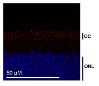
Immunofluorescence Analysis: A representative lot detected CEP290 in ciliated sensory cells (Rachel, R.A., et. al. (2012). J Clin Invest. 122(4):1233-45) and WT mouse retina (Chang, B., et. al. (2006). Hum Mol Genet. 15(11):1847-57).

Immunofluorescence Analysis: 1:500-1:2,000 dilution from a representative lot detected CEP290 in frozen mouse retina (Courtesy of Dr. Tiansen at NIH).

Immunocytochemistry Analysis: A representative lot detected CEP290 in IMCD-3 cells (Chang, B., et. al. (2006). Hum Mol Genet. 15(11):1847-57).
Biological Information
ImmunogenHis-tagged recombinant fragment corresponding to 80 amino acids from the C-terminal half of mouse Centrosomal protein of 290 kDa.
Epitopeunknown
ConcentrationPlease refer to lot specific datasheet.
HostRabbit
SpecificityThis polyclonal antibody detects centrosomal protein of 290 kDa in HEK293 cells.
Species Reactivity
  • Human
  • Rat
  • Mouse
Species Reactivity NoteHuman, Mouse, Rat.
Antibody TypePolyclonal Antibody
Entrez Gene Number
Gene Symbol
  • Cep290
  • Kiaa0373
  • Nphp6
Purification MethodAffinity Purified
UniProt Number
Molecular Weight~260 kDa observed; 289.08 kDa calculated. Uncharacterized bands may be observed in some lysate(s).
Physicochemical Information
Dimensions
Materials Information
Toxicological Information
Safety Information according to GHS
Safety Information
Product Usage Statements
Quality AssuranceEvaluated by Western Blotting in HEK293 cell lysate.

Western Blotting Analysis: 1:500 dilution of this antibody detected CEP290 in 10 µg of HEK293 cell lysate.
Usage Statement
  • Unless otherwise stated in our catalog or other company documentation accompanying the product(s), our products are intended for research use only and are not to be used for any other purpose, which includes but is not limited to, unauthorized commercial uses, in vitro diagnostic uses, ex vivo or in vivo therapeutic uses or any type of consumption or application to humans or animals.
Storage and Shipping Information
Storage ConditionsStable for 1 year at -20°C from date of receipt.

Handling Recommendations: Upon receipt and prior to removing the cap, centrifuge the vial and gently mix the solution. Aliquot into microcentrifuge tubes and store at -20°C. Avoid repeated freeze/thaw cycles, which may damage IgG and affect product performance.
Packaging Information
Material Size100 µL
Transport Information
Supplemental Information
Specifications
Global Trade Item Number
Catalogue Number GTIN
ABN1710 04054839153310