Millipore Sigma Vibrant Logo
Attention: We have moved. Merck Millipore products are no longer available for purchase on MerckMillipore.com.Learn More

NA61 Anti-BrdU (Ab-3) Mouse mAb (Mobu-1)

NA61
Purchase on Sigma-Aldrich

Overview

Replacement Information

Key Spec Table

Species ReactivityHostAntibody Type
A Broad Range Of SpeciesMMonoclonal Antibody

Products

Catalogue NumberPackaging Qty/Pack
NA61-100UG Sklená flaša 100 μg
Description
OverviewRecognizes BrdU (bromodeoxyuridine). Use of this antibody first requires biosynthetic labeling of the target cells with bromodeoxyuridine. This is the preferred BrdU antibody for staining.
Catalogue NumberNA61
Brand Family Calbiochem®
SynonymsAnti-Bromodeoxyuridine
Application Data
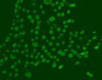
Detection of BrdU-labeled DNA by immunocytochemistry. Sample: A431 cells. Primary antibody: Anti-BrdU (Ab-3) Mouse mAb (Mobu-1) (Cat. No. NA61) (1 µg/ml). Detection: fluorescence.
References
ReferencesGratzner, H.G. 1982. Science 218, 474.
Gratzner, H.G. and Leif, R.C. 1981. Cytometry 1, 385.
Gratzner, H.G. et al. 1975. Exp. Cell Res. 95, 88.
Product Information
FormLiquid
FormulationIn 50 mM sodium phosphate buffer, 0.2% gelatin, pH 7.5.
Negative controlUnlabeled cells
Positive controlBrdU labeled DNA
Preservative≤0.1% sodium azide
Quality LevelMQ100
Applications
Application ReferencesOriginal Clone Schutte, B. et al. 1987. J. Histochem. Cytochem. 35, 1343.
Key Applications Frozen Sections
Immunocytochemistry
Not Western Blot
Paraffin Sections
Application NotesFrozen Sections (2 µg/ml)
Immunocytochemistry (1 µg/ml)
Paraffin Sections (2.5 µg/ml, trysin pre-treatment required)
Immunoblotting (not recommended)
Application CommentsTo enable antibody binding to the incorporated BrdU, cells must be fixed, permeabilized, and the DNA denatured. This is done in one step by treatment with Fixative/Denaturing Solution (70% EtOH, 0.1 N NaOH). Use of this antibody first requires biosynthetic labeling of the target cells with bromodeoxyuridine (Cat. No. 203806). Cultured cells can be pulse labeled with 10 µM bromodeoxyuridine for 30 minutes. For whole tissues, animals can be injected with 50 mg BrdU/kg body weight, sacrificed 1 h later, and the target organ(s) removed. Antibody should be titrated for optimal results in individual system.
Biological Information
ImmunogenBrdU-labeled DNA
CloneMobu-1
HostMouse
IsotypeIgG₁
Species Reactivity
  • A Broad Range Of Species
Antibody TypeMonoclonal Antibody
Concentration Label Please refer to vial label for lot-specific concentration
Physicochemical Information
Dimensions
Materials Information
Toxicological Information
Safety Information according to GHS
Safety Information
Product Usage Statements
Storage and Shipping Information
Ship Code Blue Ice Only
Toxicity Standard Handling
Storage +2°C to +8°C
Do not freeze Yes
Packaging Information
Transport Information
Supplemental Information
Specifications
Global Trade Item Number
Catalogue Number GTIN
NA61-100UG 04055977227604

Documentation

Anti-BrdU (Ab-3) Mouse mAb (Mobu-1) MSDS

Title

Safety Data Sheet (SDS) 

Anti-BrdU (Ab-3) Mouse mAb (Mobu-1) Certificates of Analysis

TitleLot Number
NA61

References

Reference overview
Gratzner, H.G. 1982. Science 218, 474.
Gratzner, H.G. and Leif, R.C. 1981. Cytometry 1, 385.
Gratzner, H.G. et al. 1975. Exp. Cell Res. 95, 88.

Brochure

Title
Caspases and other Apoptosis Related Tools Brochure
Data Sheet

Note that this data sheet is not lot-specific and is representative of the current specifications for this product. Please consult the vial label and the certificate of analysis for information on specific lots. Also note that shipping conditions may differ from storage conditions.

Revision05-September-2007 RFH
SynonymsAnti-Bromodeoxyuridine
ApplicationFrozen Sections (2 µg/ml)
Immunocytochemistry (1 µg/ml)
Paraffin Sections (2.5 µg/ml, trysin pre-treatment required)
Immunoblotting (not recommended)
Application Data
Detection of BrdU-labeled DNA by immunocytochemistry. Sample: A431 cells. Primary antibody: Anti-BrdU (Ab-3) Mouse mAb (Mobu-1) (Cat. No. NA61) (1 µg/ml). Detection: fluorescence.
DescriptionPurified mouse monoclonal antibody generated by immunizing mice with the specified immunogen and fusing splenocytes with P3-X63-Ag8-653 mouse myeloma cells (see application references). Recognizes BrdU (bromodeoxyuridine).
BackgroundProliferating cells are usually detected by incorporation of [3H]-thymidine into DNA and quantitation by autoradiography or scintillation counting. Alternatively, bromodeoxyuridine (BrdU), an analog of thymidine, can be incorporated specifically into the DNA of replicating cells and the extent of incorporation can be detected using antibodies directed against the incorporated analog. The proportion of cells actively proliferating can then be determined by immunofluorescence or flow cytometry as well as immunohistochemistry (which has been done with tumors, tissues, and blood cell elements).
HostMouse
ImmunogenBrdU-labeled DNA
CloneMobu-1
IsotypeIgG₁
Speciesa broad range of species
Positive controlBrdU labeled DNA
Negative controlUnlabeled cells
FormLiquid
FormulationIn 50 mM sodium phosphate buffer, 0.2% gelatin, pH 7.5.
Concentration Label Please refer to vial label for lot-specific concentration
Preservative≤0.1% sodium azide
CommentsTo enable antibody binding to the incorporated BrdU, cells must be fixed, permeabilized, and the DNA denatured. This is done in one step by treatment with Fixative/Denaturing Solution (70% EtOH, 0.1 N NaOH). Use of this antibody first requires biosynthetic labeling of the target cells with bromodeoxyuridine (Cat. No. 203806). Cultured cells can be pulse labeled with 10 µM bromodeoxyuridine for 30 minutes. For whole tissues, animals can be injected with 50 mg BrdU/kg body weight, sacrificed 1 h later, and the target organ(s) removed. Antibody should be titrated for optimal results in individual system.
Storage +2°C to +8°C
Do Not Freeze Yes
Toxicity Standard Handling
ReferencesGratzner, H.G. 1982. Science 218, 474.
Gratzner, H.G. and Leif, R.C. 1981. Cytometry 1, 385.
Gratzner, H.G. et al. 1975. Exp. Cell Res. 95, 88.
Application referencesOriginal Clone Schutte, B. et al. 1987. J. Histochem. Cytochem. 35, 1343.