Epigenetic analysis reveals a euchromatic configuration in the FMR1 unmethylated full mutations.
Tabolacci, E; Moscato, U; Zalfa, F; Bagni, C; Chiurazzi, P; Neri, G
European journal of human genetics : EJHG
16
1487-98
2008
Show Abstract
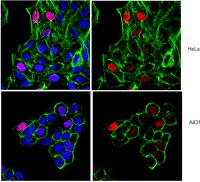
Fragile X syndrome (FXS) is caused by the expansion of a CGG repeat in the 5'UTR of the FMR1 gene and the subsequent methylation of all CpG sites in the promoter region. We recently identified, in unrelated FXS families, two rare males with an unmethylated full mutation, that is, with an expanded CGG repeat (greater than 200 triplets) lacking the typical CpG methylation in the FMR1 promoter. These individuals are not mentally retarded and do not appear to be mosaic for premutation or methylated full mutation alleles. We established lymphoblastoid and fibroblast cell lines that showed essentially normal levels of the FMR1-mRNA but reduced translational efficiency of the corresponding mRNA. Epigenetic analysis of the FMR1 gene demonstrated the lack of DNA methylation and a methylation pattern of lysines 4 and 27 on histone H3 similar to that of normal controls, in accordance with normal transcription levels and consistent with a euchromatic configuration. On the other hand, histone H3/H4 acetylation and lysine 9 methylation on histone H3 were similar to those of typical FXS cell lines, suggesting that these epigenetic changes are not sufficient for FMR1 gene inactivation. These findings demonstrate remarkable consistency and suggest a common genetic mechanism causing this rare FMR1 epigenotype. The discovery of such a mechanism may be important in view of therapeutic attempts to convert methylated into unmethylated full mutations, restoring the expression of the FMR1 gene. | Chromatin Immunoprecipitation (ChIP) | Human | 18628788
 |
Chromatin remodeling at the Th2 cytokine gene loci in human type 2 helper T cells.
Takaaki Kaneko,Hiroyuki Hosokawa,Masakatsu Yamashita,Chrong-Reen Wang,Akihiro Hasegawa,Motoko Y Kimura,Masayuki Kitajiama,Fumio Kimura,Masaru Miyazaki,Toshinori Nakayama
Molecular immunology
44
2007
Show Abstract
The differentiation of mouse naïve CD4 T cells into type 2 helper (Th2) cells is accompanied by chromatin remodeling at the nucleosomes associated with the IL-4, IL-13 and IL-5 genes. However, little is known about how chromatin remodeling of these Th2 cytokine gene loci occurs in human Th2 cells. We herein established an in vitro culture system in which both Th1 and Th2 cells are efficiently differentiated from human peripheral blood naïve CD4 T cells. This system allowed us to investigate the chromatin status at the Th2 cytokine gene loci and the IFNgamma locus in human Th2 and Th1 cells, respectively. In typical individuals, the chromatin remodeling indicated by the induction of hyper-acetylation of histone H3 lysine 9 and hyper-methylation of histone H3 lysine 4 was induced at the whole Th2 cytokine gene loci in developing Th2 cells. We more precisely assessed the methylation status of histone H3 lysine 4 at the Th2 cytokine gene loci (IL-5 exon 3, IL-5 promoter, IL-5/RAD50 intergenic region, RAD50 promoter, CGRE, CNS1, IL-13 promoter, IL-4 promoter, and VA enhancer regions) and the IFNgamma locus in developing Th1 and Th2 cells prepared from 20 healthy volunteers. Th2-cell specific chromatin remodeling was induced at most of the Th2 cytokine gene loci. In parallel with the induction of chromatin remodeling, GATA3 mRNA was preferentially expressed in developing Th2 cells, whereas T-bet, HLX and ROG mRNA was selectively expressed in developing Th1 cells. | | | 17166591
|
Epigenetic patterns of the retinoic acid receptor beta2 promoter in retinoic acid-resistant thyroid cancer cells.
Cras, A, et al.
Oncogene, 26: 4018-24 (2007)
2007
Show Abstract
Treatment with retinoic acid (RA) is effective to restore radioactive iodine uptake in metastases of a small fraction of thyroid cancer patients. In order to find predictive markers of response, we took advantage of two thyroid cancer cell lines, FTC133 and FTC238, with low RA-receptor (RAR)beta expression but differing in their response to RA. We report that in both cell lines, RA signalling pathways are functional, as transactivation of an exogenous RARbeta2 promoter is effective in the presence of pharmacological concentrations of all-trans RA, and enhanced in RA-resistant FTC238 cells after ectopical expression of RARbeta, suggesting a defective endogenous RARbeta2 promoter in these cells. Further analyses show that whereas the RARbeta2 promoter is in an unmethylated permissive status in both FTC133 and FTC238 cells, it failed to be associated with acetylated forms of histones H3 or H4 in FTC238 cells upon RA treatment. Incubation with a histone deacetylase inhibitor, alone or in combination with RA, restored histone acetylation levels and reactivated RARbeta and differentiation marker Na+/I- symporter gene expression. Thus, histone modification patterns may explain RA-refractoriness in differentiated thyroid cancer patients and suggest a potential benefit of combined transcriptional and differentiation therapies. | | | 17213810
|
Inhibition of histone deacetylase activity induces developmental plasticity in oligodendrocyte precursor cells.
Costas A Lyssiotis,John Walker,Chunlei Wu,Toru Kondo,Peter G Schultz,Xu Wu
Proceedings of the National Academy of Sciences of the United States of America
104
2007
Show Abstract
Recently, it was demonstrated that lineage-committed oligodendrocyte precursor cells (OPCs) can be converted to multipotent neural stem-like cells, capable of generating both neurons and glia after exposure to bone morphogenetic proteins. In an effort to understand and control the developmental plasticity of OPCs, we developed a high-throughput screen to identify novel chemical inducers of OPC reprogramming. Using this system, we discovered that inhibition of histone deacetylase (HDAC) activity in OPCs acts as a priming event in the induction of developmental plasticity, thereby expanding the differentiation potential to include the neuronal lineage. This conversion was found to be mediated, in part, through reactivation of sox2 and was highly reproducible at the clonal level. Further, genome-wide expression analysis demonstrated that HDAC inhibitor treatment activated sox2 and 12 other genes that identify or maintain the neural stem cell state while simultaneously silencing a large group of oligodendrocyte lineage-specific genes. This series of experiments demonstrates that global histone acetylation, induced by HDAC inhibition, can partially reverse the lineage restriction of OPCs, thereby inducing developmental plasticity. Full Text Article | | | 17855562
|
Critical YxKxHxxxRP motif in the C-terminal region of GATA3 for its DNA binding and function.
Ryo Shinnakasu, Masakatsu Yamashita, Kenta Shinoda, Yusuke Endo, Hiroyuki Hosokawa, Akihiro Hasegawa, Shinji Ikemizu, Toshinori Nakayama
Journal of immunology (Baltimore, Md. : 1950)
177
5801-10
2005
Show Abstract
A zinc finger transcription factor, GATA3, plays an essential role in the development of T cells and the functional differentiation into type 2 Th cells. Two transactivation domains and two zinc finger regions are known to be important for the GATA3 function, whereas the role for other regions remains unclear. In this study we demonstrated that a conserved YxKxHxxxRP motif (aa 345-354) adjacent to the C-terminal zinc finger domain of GATA3 plays a critical in its DNA binding and functions, including transcriptional activity, the ability to induce chromatin remodeling of the Th2 cytokine gene loci, and Th2 cell differentiation. A single point mutation of the key amino acid (Y, K, H, R, and P) in the motif abrogated GATA3 functions. A computer simulation analysis based on the solution structure of the chicken GATA1/DNA complex supported the importance of this motif in GATA3 DNA binding. Thus, we identified a novel conserved YxKxHxxxRP motif adjacent to the C-terminal zinc finger domain of GATA3 that is indispensable for GATA3 DNA binding and functions. | | | 17056504
|
Ras-ERK MAPK cascade regulates GATA3 stability and Th2 differentiation through ubiquitin-proteasome pathway.
Masakatsu Yamashita, Ryo Shinnakasu, Hikari Asou, Motoko Kimura, Akihiro Hasegawa, Kahoko Hashimoto, Naoya Hatano, Masato Ogata, Toshinori Nakayama
The Journal of biological chemistry
280
29409-19
2004
Show Abstract
Differentiation of naive CD4 T cells into Th2 cells requires protein expression of GATA3. Interleukin-4 induces STAT6 activation and subsequent GATA3 transcription. Little is known, however, on how T cell receptor-mediated signaling regulates GATA3 and Th2 cell differentiation. Here we demonstrated that T cell receptor-mediated activation of the Ras-ERK MAPK cascade stabilizes GATA3 protein in developing Th2 cells through the inhibition of the ubiquitin-proteasome pathway. Mdm2 was associated with GATA3 and induced ubiquitination on GATA3, suggesting its role as a ubiquitin-protein isopeptide ligase for GATA3 ubiquitination. Thus, the Ras-ERK MAPK cascade controls GATA3 protein stability by a post-transcriptional mechanism and facilitates GATA3-mediated chromatin remodeling at Th2 cytokine gene loci leading to successful Th2 cell differentiation. | | | 15975924
|
Differential expression of IFN regulatory factor 4 gene in human monocyte-derived dendritic cells and macrophages.
Anne Lehtonen, Ville Veckman, Tuomas Nikula, Riitta Lahesmaa, Leena Kinnunen, Sampsa Matikainen, Ilkka Julkunen
Journal of immunology (Baltimore, Md. : 1950)
175
6570-9
2004
Show Abstract
In vitro human monocyte differentiation to macrophages or dendritic cells (DCs) is driven by GM-CSF or GM-CSF and IL-4, respectively. IFN regulatory factors (IRFs), especially IRF1 and IRF8, are known to play essential roles in the development and functions of macrophages and DCs. In the present study, we performed cDNA microarray and Northern blot analyses to characterize changes in gene expression of selected genes during cytokine-stimulated differentiation of human monocytes to macrophages or DCs. The results show that the expression of IRF4 mRNA, but not of other IRFs, was specifically up-regulated during DC differentiation. No differences in IRF4 promoter histone acetylation could be found between macrophages and DCs, suggesting that the gene locus was accessible for transcription in both cell types. Computer analysis of the human IRF4 promoter revealed several putative STAT and NF-kappaB binding sites, as well as an IRF/Ets binding site. These sites were found to be functional in transcription factor-binding and chromatin immunoprecipitation experiments. Interestingly, Stat4 and NF-kappaB p50 and p65 mRNAs were expressed at higher levels in DCs as compared with macrophages, and enhanced binding of these factors to their respective IRF4 promoter elements was found in DCs. IRF4, together with PU.1, was also found to bind to the IRF/Ets response element in the IRF4 promoter, suggesting that IRF4 protein provides a positive feedback signal for its own gene expression in DCs. Our results suggest that IRF4 is likely to play an important role in myeloid DC differentiation and gene regulatory functions. | | | 16272311
|
CD28 costimulation controls histone hyperacetylation of the interleukin 5 gene locus in developing th2 cells.
Masamichi Inami, Masakatsu Yamashita, Yoshiyuki Tenda, Akihiro Hasegawa, Motoko Kimura, Kahoko Hashimoto, Nobuo Seki, Masaru Taniguchi, Toshinori Nakayama
The Journal of biological chemistry
279
23123-33
2004
Show Abstract
Interleukin 5 (IL-5) plays a unique role in allergic inflammatory responses, and the understanding of molecular mechanisms underlying the generation of IL-5-producing cells is crucial for the regulation of allergic disorders. Differentiation of naive CD4 T cells into type-2 helper (Th2) cells is accompanied by chromatin remodeling including hyperacetylation of histones H3 and H4 in the nucleosomes associated with the IL-4, IL-13, and IL-5 genes. Histone hyperacetylation of the IL-5 gene displayed a delayed kinetics compared with that of the IL-4 and IL-13 genes, suggesting a distinct remodeling mechanism for the IL-5-gene locus. Here we studied the role of CD28 costimulation in the generation of IL-5-producing cells and the histone hyperacetylation of the IL-5 gene locus. CD28-costimulation selectively enhanced histone hyperacetylation of the IL-5 gene locus that appeared to be mediated through NF-kappaB activation and subsequent up-regulation of GATA3. The CD28 costimulation-sensitive histone hyperacetylation spanned almost the entire intergenic region between the IL-5 and RAD50 accompanied with intergenic transcript. Thus, this is the first demonstration that CD28 costimulation controls a chromatin-remodeling process during Th2 cell differentiation. | | | 15039422
|
Essential role of GATA3 for the maintenance of type 2 helper T (Th2) cytokine production and chromatin remodeling at the Th2 cytokine gene loci.
Masakatsu Yamashita, Maki Ukai-Tadenuma, Takeshi Miyamoto, Kaoru Sugaya, Hiroyuki Hosokawa, Akihiro Hasegawa, Motoko Kimura, Masaru Taniguchi, James DeGregori, Toshinori Nakayama
The Journal of biological chemistry
279
26983-90
2004
Show Abstract
GATA3 expression is essential for type-2 helper T (Th2) cell differentiation. GATA3-mediated chromatin remodeling at the Th2 cytokine gene loci, including Th2-specific long range histone hyperacetylation of the interleukin (IL)-13/IL-4 gene loci, occurs in developing Th2 cells. However, little is known about the role of GATA3, if any, in the maintenance of established remodeled chromatin at the Th2 cytokine gene loci. Here, we established a Cre/LoxP-based site-specific recombination system in cultured CD4 T cells using a unique adenovirus-mediated gene transfer technique. This system allowed us to investigate the effect of loss of GATA3 expression in in vitro differentiated Th2 cells. After ablation of GATA3, we detected reduced production of all Th2 cytokines, increased DNA methylation at the IL-4 gene locus, and decreased histone hyperacetylation at the IL-5 gene locus but not significantly so at the IL-13/IL-4 gene loci. Thus, GATA3 plays important roles in the maintenance of the Th2 phenotype and continuous chromatin remodeling of the specific Th2 cytokine gene locus through cell division. | | | 15087456
|
Cyclin D1 activation in B-cell malignancy: association with changes in histone acetylation, DNA methylation, and RNA polymerase II binding to both promoter and distal sequences.
Hui Liu, Jin Wang, Elliot M Epner
Blood
104
2505-13
2004
Show Abstract
Cyclin D1 expression is deregulated by chromosome translocation in mantle cell lymphoma and a subset of multiple myeloma. The molecular mechanisms involved in long-distance gene deregulation remain obscure, although changes in acetylated histones and methylated CpG dinucleotides may be important. The patterns of DNA methylation and histone acetylation were determined at the cyclin D1 locus on chromosome 11q13 in B-cell malignancies. The cyclin D1 promoter was hypomethylated and hyperacetylated in expressing cell lines and patient samples, and methylated and hypoacetylated in nonexpressing cell lines. Domains of hyperacetylated histones and hypomethylated DNA extended over 120 kb upstream of the cyclin D1 gene. Interestingly, hypomethylated DNA and hyperacetylated histones were also located at the cyclin D1 promoter but not the upstream major translocation cluster region in cyclin D1-nonexpressing, nontumorigenic B and T cells. RNA polymerase II binding was demonstrated both at the cyclin D1 promoter and 3' immunoglobulin heavy-chain regulatory regions only in malignant B-cell lines with deregulated cyclin D1 expression. Our results suggest a model where RNA polymerase II bound at IgH regulatory sequences can activate the cyclin D1 promoter by either long-range polymerase transfer or tracking. | | | 15226187
|