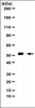
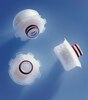
Millipore Sigma Vibrant Logo
Attention: We have moved. Merck Millipore products are no longer available for purchase on MerckMillipore.com.Learn More

281753 DAB Substrate Buffer

281753
  
Purchase on Sigma-Aldrich

Přehled

Replacement Information
Description
Overview

This product has been discontinued.


Peroxidase substrate buffer supplied as a 10X concentrate (pH 7.6 when diluted). A diluent for Cat. No. 281751.
Catalogue Number281753
Brand Family Calbiochem®
References
Product Information
FormClear liquid
PreservativeNone
Applications
Application NotesDesigned for use with DAB, Tetrahydrochloride, Cat. No. 281751.
Application CommentsStable at 4°C, 18-26°C, and 37°C. pH = 7.6 (after dilution). Functionality check performed on final product using immunohistological staining.

Suggested Procedure for Immunohistochemical Staining Using DAB:

1. Complete all required incubations with antibodies and HRP-labeled probes, and all necessary wash steps.
2. Prepare 1X DAB Substrate Buffer: Add 9 parts distilled water to 1 part 10X DAB Substrate Buffer (Cat. No. 281753). Mix well. [Note: 1 M Tris-HCl, pH 7.6 may be substituted for Cat. No. 281753, such that the final (1X) concentration is 100 mM Tris-HCl, pH 7.6.
3. Prepare 1X DAB Substrate Solution: Add 1 part 50X DAB Concentrate and 1 part 0.5% H2O2 to 50 parts 1X DAB Substrate Buffer (prepared in Step 2). Mix well and protect from light.
4. Following the final wash, completely cover tissue sections with the 1X DAB Substrate Solution (prepared in Step 3) and incubate 5-15 min at room temperature. Note: Variables associated with assay conditions will dictate the proper reaction time.
5. After reaction is complete, wash tissue sections thoroughly in distilled water.
6. Counterstain with hematoxylin if desired.
7. Dehydrate in graded alcohols, xylene, or xylene substitutes.
8. Mount tissue sections using xylene-based mounting media.

Suggested Procedure for Immunoblot Staining with DAB:

1. Complete all required incubations with antibodies and HRP-labeled probes, and all necessary wash steps.
2. Prepare 1X DAB Substrate Buffer: Add 9 parts distilled water to 1 part 10X DAB Substrate Buffer (Cat. No. 281753). Mix well. [Note: 1 M Tris-HCl, pH 7.6 may be substituted for Cat. No. 281753, such that the final (1X) concentration is 100 mM Tris-HCl, pH 7.6.]
3. Prepare 1X DAB Substrate Solution: Add 1 part 50X DAB Concentrate and 1 part 0.5% H2O2 to 50 parts 1X DAB Substrate Buffer (prepared in Step 2). Mix well and protect from light.
4. Following the final wash, completely cover membranes with 1X DAB Substrate Solution (prepared in Step 3). Incubate 5-30 min at room temperature.
Note: Variables associated with assay conditions will dictate the proper reaction time.
5. After reaction is complete, wash membranes thoroughly in distilled water.
6. Air-dry membranes and store protected from light.
Biological Information
Physicochemical Information
Dimensions
Materials Information
Toxicological Information
Safety Information according to GHS
Safety Information
Product Usage Statements
Storage and Shipping Information
Ship Code Ambient Temperature Only
Toxicity Standard Handling
Storage +2°C to +8°C
Do not freeze Ok to freeze
Packaging Information
Transport Information
Supplemental Information
Specifications
Global Trade Item Number
Katalogové číslo GTIN
281753 0

Documentation

DAB Substrate Buffer Certificates of Analysis

TitleLot Number
281753
Data Sheet

Note that this data sheet is not lot-specific and is representative of the current specifications for this product. Please consult the vial label and the certificate of analysis for information on specific lots. Also note that shipping conditions may differ from storage conditions.

Revision21-August-2007 JSW
ApplicationDesigned for use with DAB, Tetrahydrochloride, Cat. No. 281751.
DescriptionA buffer diluent for use with DAB, a precipitable peroxidase substrate for immunoblotting and immunohistochemical staining techniques. Supplied as 50 ml of a 10X concentrate.
BackgroundSince first introduced by Graham and Karnovsky numerous procedures for the use of DAB for detection of horseradish peroxidase (HRP)-labeled probes in histochemistry, immunohistochemistry, western blots and dot blots have been described. In the presence of horseradish peroxidase and hydrogen peroxide, DAB is oxidized to a brown polymer that is insoluble in most organic solvents. Thus, xylene-based mounting media may be used for immunohistochemical applications. Sites of HRP activity on tissue sections and blots appear as brown-orange deposits. The color can be modified and intensified by treatment with salts of silver, copper, nickel, cobalt and osmium. This DAB substrate preparation is a stable, convenient, liquid concentrate in a proprietary solvent. The stabilization system prevents formation of partially oxidized DAB, thus eliminating the nonspecific binding to other heme-containing proteins so often observed with powdered DAB preparations. The concentrate can be diluted in appropriate peroxide-containing buffers, providing the researcher with the capability of formulating any of the numerous published DAB reaction systems.
FormClear liquid
PreservativeNone
CommentsStable at 4°C, 18-26°C, and 37°C. pH = 7.6 (after dilution). Functionality check performed on final product using immunohistological staining.

Suggested Procedure for Immunohistochemical Staining Using DAB:

1. Complete all required incubations with antibodies and HRP-labeled probes, and all necessary wash steps.
2. Prepare 1X DAB Substrate Buffer: Add 9 parts distilled water to 1 part 10X DAB Substrate Buffer (Cat. No. 281753). Mix well. [Note: 1 M Tris-HCl, pH 7.6 may be substituted for Cat. No. 281753, such that the final (1X) concentration is 100 mM Tris-HCl, pH 7.6.
3. Prepare 1X DAB Substrate Solution: Add 1 part 50X DAB Concentrate and 1 part 0.5% H2O2 to 50 parts 1X DAB Substrate Buffer (prepared in Step 2). Mix well and protect from light.
4. Following the final wash, completely cover tissue sections with the 1X DAB Substrate Solution (prepared in Step 3) and incubate 5-15 min at room temperature. Note: Variables associated with assay conditions will dictate the proper reaction time.
5. After reaction is complete, wash tissue sections thoroughly in distilled water.
6. Counterstain with hematoxylin if desired.
7. Dehydrate in graded alcohols, xylene, or xylene substitutes.
8. Mount tissue sections using xylene-based mounting media.

Suggested Procedure for Immunoblot Staining with DAB:

1. Complete all required incubations with antibodies and HRP-labeled probes, and all necessary wash steps.
2. Prepare 1X DAB Substrate Buffer: Add 9 parts distilled water to 1 part 10X DAB Substrate Buffer (Cat. No. 281753). Mix well. [Note: 1 M Tris-HCl, pH 7.6 may be substituted for Cat. No. 281753, such that the final (1X) concentration is 100 mM Tris-HCl, pH 7.6.]
3. Prepare 1X DAB Substrate Solution: Add 1 part 50X DAB Concentrate and 1 part 0.5% H2O2 to 50 parts 1X DAB Substrate Buffer (prepared in Step 2). Mix well and protect from light.
4. Following the final wash, completely cover membranes with 1X DAB Substrate Solution (prepared in Step 3). Incubate 5-30 min at room temperature.
Note: Variables associated with assay conditions will dictate the proper reaction time.
5. After reaction is complete, wash membranes thoroughly in distilled water.
6. Air-dry membranes and store protected from light.
Storage +2°C to +8°C
Do Not Freeze Ok to freeze
Toxicity Standard Handling