csrnp1a is necessary for the development of primitive hematopoiesis progenitors in zebrafish.
Espina, J; Feijóo, CG; Solís, C; Glavic, A
PloS one
8
e53858
2013
Zobrazit abstrakt
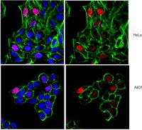
The CSRNP (cystein-serine-rich nuclear protein) transcription factors are conserved from Drosophila to human. Functional studies in mice, through knockout for each of their paralogs, have resulted insufficient to elucidate the function of this family of proteins in vertebrate development. Previously, we described the function of the zebrafish ortholog, Csnrp1/Axud1, showing its essential role in the survival and proliferation of cephalic progenitors. To extend our understanding of this family, we have studied the function of its paralog csrnp1a. Our results show that csrnp1a is expressed from 0 hpf, until larval stages, particularly in cephalic territories and in the intermediate cell mass (ICM). Using morpholinos in wild type and transgenic lines we observed that Csrnp1a knockdown generates a mild reduction in head size and a depletion of blood cells in circulation. This was combined with in situ hybridizations to analyze the expression of different mesodermal and primitive hematopoiesis markers. Morphant embryos have impaired blood formation without disruption of mesoderm specification, angiogenesis or heart development. The reduction of circulating blood cells occurs at the hematopoietic progenitor level, affecting both the erythroid and myeloid lineages. In addition, cell proliferation was also altered in hematopoietic anterior sites, specifically in spi1 expression domain. These and previous observations suggest an important role of Csnrps transcription factors in progenitor biology, both in the neural and hematopoietic linages. | Immunohistochemistry | 23326522
 |
Lasonolide A, a potent and reversible inducer of chromosome condensation.
Zhang, YW; Ghosh, AK; Pommier, Y
Cell cycle (Georgetown, Tex.)
11
4424-35
2011
Zobrazit abstrakt
Lasonolide A (LSA) is a natural product with high and selective cytotoxicity against mesenchymal cancer cells, including leukemia, melanomas and glioblastomas. Here, we reveal that LSA induces rapid and reversible premature chromosome condensation (PCC) associated with cell detachment, plasma membrane smoothening and actin reorganization. PCC is induced at all phases of the cell cycle in proliferative cells as well as in circulating human lymphocytes in G 0. It is independent of Cdk1 signaling, associated with cyclin B downregulation and induced in cells at LSA concentrations that are three orders of magnitude lower than those required to block phosphatases 1 and 2A in vitro. At the epigenetic level, LSA-induced PCC is coupled with histone H3 and H1 hyperphosphorylation and deacetylation. Treatment with SAHA reduced LSA-induced PCC, implicating histone deacetylation as one of the PCC effector mechanisms. In addition, PCC is coupled with topoisomerase II (Top2) and Aurora A hyperphosphorylation and activation. Inhibition of Top2 or Aurora A partially blocked LSA-induced PCC. Our findings demonstrate the profound epigenetic alterations induced by LSA and the potential of LSA as a new cytogenetic tool. Based on the unique cellular effects of LSA, further studies are warranted to uncover the cellular target of lasonolide A ("TOL"). | Western Blotting | 23159859
|
AtHaspin phosphorylates histone H3 at threonine 3 during mitosis and contributes to embryonic patterning in Arabidopsis.
Raheleh Karimi Ashtiyani,Ali Mohammad Banaei Moghaddam,Veit Schubert,Twan Rutten,Jörg Fuchs,Dmitri Demidov,Frank R Blattner,Andreas Houben
The Plant journal : for cell and molecular biology
68
2010
Zobrazit abstrakt
Post-translational histone modifications regulate many aspects of chromosome activity. Threonine 3 of histone H3 is highly conserved, but the significance of its phosphorylation is unclear, and the identity of the corresponding kinase in plants is unknown. Therefore, we characterized the candidate kinase in Arabidopsis thaliana, called AtHaspin. Recombinant AtHaspin in vitro phosphorylates histone H3 at threonine 3. Reduction of H3 threonine 3 phosphorylation level and reduced chromatin condensation in interphase nuclei by AtHaspin RNAi supports the proposition that this kinase is involved in histone H3 phosphorylation in vivo in mitotic cells. In addition, we provide a developmental function for a Haspin kinase. At the whole plant level, altered expression of the kinase induced pleiotropic phenotypes with defects in floral organs and vascular tissue. It reduced fertility and modified adventitious shoot apical meristems that then gave rise to plants with multi-rosettes and multi-shoots. Haspin mutant embryos frequently showed alteration in division plane orientation that could be traced back to the earliest divisions of embryo development, thus Haspin contributes to embryonic patterning. | | 21749502
|
PP1/Repo-Man Dephosphorylates Mitotic Histone H3 at T3 and Regulates Chromosomal Aurora B Targeting.
Qian, Junbin, et al.
Curr. Biol., 21: 766-73 (2011)
2010
Zobrazit abstrakt
The transient mitotic histone H3 phosphorylation by various protein kinases regulates chromosome condensation and segregation, but the counteracting phosphatases have been poorly characterized [1-8]. We show here that PP1γ is the major histone H3 phosphatase acting on the mitotically phosphorylated (ph) residues H3T3ph, H3S10ph, H3T11ph, and H3S28ph. In addition, we identify Repo-Man, a chromosome-bound interactor of PP1γ [9], as a selective regulator of H3T3ph and H3T11ph dephosphorylation. Repo-Man promotes H3T11ph dephosphorylation by an indirect mechanism but directly and specifically targets H3T3ph for dephosphorylation by associated PP1γ. The PP1γ/Repo-Man complex opposes the protein kinase Haspin-mediated spreading of H3T3ph to the chromosome arms until metaphase and catalyzes the net dephosphorylation of H3T3ph at the end of mitosis. Consistent with these findings, Repo-Man modulates in a PP1-dependent manner the H3T3ph-regulated chromosomal targeting of Aurora kinase B and its substrate MCAK. Our study defines a novel mechanism by which PP1 counteracts Aurora B. | | 21514157
|
The human CDK8 subcomplex is a histone kinase that requires Med12 for activity and can function independently of mediator.
Knuesel, MT; Meyer, KD; Donner, AJ; Espinosa, JM; Taatjes, DJ
Molecular and cellular biology
29
650-61
2009
Zobrazit abstrakt
The four proteins CDK8, cyclin C, Med12, and Med13 can associate with Mediator and are presumed to form a stable "CDK8 subcomplex" in cells. We describe here the isolation and enzymatic activity of the 600-kDa CDK8 subcomplex purified directly from human cells and also via recombinant expression in insect cells. Biochemical analysis of the recombinant CDK8 subcomplex identifies predicted (TFIIH and RNA polymerase II C-terminal domain [Pol II CTD]) and novel (histone H3, Med13, and CDK8 itself) substrates for the CDK8 kinase. Notably, these novel substrates appear to be metazoan-specific. Such diverse targets imply strict regulation of CDK8 kinase activity. Along these lines, we observe that Mediator itself enables CDK8 kinase activity on chromatin, and we identify Med12--but not Med13--to be essential for activating the CDK8 kinase. Moreover, mass spectrometry analysis of the endogenous CDK8 subcomplex reveals several associated factors, including GCN1L1 and the TRiC chaperonin, that may help control its biological function. In support of this, electron microscopy analysis suggests TRiC sequesters the CDK8 subcomplex and kinase assays reveal the endogenous CDK8 subcomplex--unlike the recombinant submodule--is unable to phosphorylate the Pol II CTD. Celý text článku | | 19047373
|
Conditional mutations of beta-catenin and APC reveal roles for canonical Wnt signaling in lens differentiation.
Martinez, G; Wijesinghe, M; Turner, K; Abud, HE; Taketo, MM; Noda, T; Robinson, ML; de Iongh, RU
Investigative ophthalmology & visual science
50
4794-806
2009
Zobrazit abstrakt
Previous studies indicate that the Wnt/beta-catenin-signaling pathway is active and functional during murine lens development. In this study, the consequences of constitutively activating the pathway in lens during development were investigated.To activate Wnt/beta-catenin signaling, beta-catenin (Catnb) and adenomatous polyposis coli (Apc) genes were conditionally mutated in two Cre lines that are active in whole lens (MLR10) or only in differentiated fibers (MLR39), from E13.5. Lens phenotype in mutant lenses was investigated by histology, immunohistochemistry, BrdU labeling, quantitative RT-PCR arrays, and TUNEL.Only intercrosses with MLR10 resulted in ocular phenotypes, indicating Wnt/beta-catenin signaling functions in lens epithelium and during early fiber differentiation. Mutant lenses were characterized by increased progression of epithelial cells through the cell cycle, as shown by BrdU labeling, and phosphohistone 3 and cyclin D1 labeling, and maintenance of epithelial phenotype (E-cadherin and Pax6 expression) in the fiber compartment. Fiber cell differentiation was delayed as shown by reduced expression of c-maf and beta-crystallin and delay in expression of the CDKI, p57(kip2). From E13.5, there were numerous cells undergoing apoptosis, and by E15.5, there was evidence of epithelial-mesenchymal transition with numerous cells expressing alpha-smooth muscle actin. Quantitative PCR analyses revealed large changes in expression of Wnt target genes (Lef1, Tcf7, T (Brachyury), and Ccnd1), Wnt inhibitors (Wif1, Dkk1, Nkd1, and Frzb) and also several Wnts (Wnt6, Wnt10a, Wnt8b, and Wnt11).These data indicate that the Wnt/beta-catenin pathway plays key roles in regulating proliferation of lens stem/progenitor cells during early stages of fiber cell differentiation. | | 19515997
|
The MUT9p kinase phosphorylates histone H3 threonine 3 and is necessary for heritable epigenetic silencing in Chlamydomonas.
Casas-Mollano, JA; Jeong, BR; Xu, J; Moriyama, H; Cerutti, H
Proceedings of the National Academy of Sciences of the United States of America
105
6486-91
2008
Zobrazit abstrakt
Changes in chromatin organization are emerging as key regulators in nearly every aspect of DNA-templated metabolism in eukaryotes. Histones undergo many, largely reversible, posttranslational modifications that affect chromatin structure. Some modifications, such as trimethylation of histone H3 on Lys 4 (H3K4me3), correlate with transcriptional activation, whereas others, such as methylation of histone H3 on Lys 27 (H3K27me), are associated with silent chromatin. Posttranslational histone modifications may also be involved in the inheritance of chromatin states. Histone phosphorylation has been implicated in a variety of cellular processes but, because of the dynamic nature of this modification, its potential role in long-term gene silencing has remained relatively unexplored. We report here that a Chlamydomonas reinhardtii mutant defective in a Ser/Thr protein kinase (MUT9p), which phosphorylates histones H3 and H2A, shows deficiencies in the heritable repression of transgenes and transposons. Moreover, based on chromatin immunoprecipitation analyses, phosphorylated H3T3 (H3T3ph) and monomethylated H3K4 (H3K4me1) are inversely correlated with di/trimethylated H3K4 and associate preferentially with silenced transcription units. Conversely, the loss of those marks in mutant strains correlates with the transcriptional reactivation of transgenes and transposons. Our results suggest that H3T3ph and H3K4me1 function as reinforcing epigenetic marks for the silencing of euchromatic loci in Chlamydomonas. | Western Blotting | 18420823
|
Identification and dynamics of two classes of aurora-like kinases in Arabidopsis and other plants.
Demidov, D; Van Damme, D; Geelen, D; Blattner, FR; Houben, A
The Plant cell
17
836-48
2004
Zobrazit abstrakt
Aurora-like kinases play key roles in chromosome segregation and cytokinesis in yeast, plant, and animal systems. Here, we characterize three Arabidopsis thaliana protein kinases, designated AtAurora1, AtAurora2, and AtAurora3, which share high amino acid identities with the Ser/Thr kinase domain of yeast Ipl1 and animal Auroras. Structure and expression of AtAurora1 and AtAurora2 suggest that these genes arose by a recent gene duplication, whereas the diversification of plant alpha and beta Aurora kinases predates the origin of land plants. The transcripts and proteins of all three kinases are most abundant in tissues containing dividing cells. Intracellular localization of green fluorescent protein-tagged AtAuroras revealed an AtAurora-type specific association mainly with dynamic mitotic structures, such as microtubule spindles and centromeres, and with the emerging cell plate of dividing tobacco (Nicotiana tabacum) BY-2 cells. Immunolabeling using AtAurora antibodies yielded specific signals at the centromeres that are coincident with histone H3 that is phosphorylated at Ser position10 during mitosis. An in vitro kinase assay demonstrated that AtAurora1 preferentially phosphorylates histone H3 at Ser 10 but not at Ser 28 or Thr 3, 11, and 32. The phylogenetic analysis of available Aurora sequences from different eukaryotic origins suggests that, although a plant Aurora gene has been duplicated early in the evolution of plants, the paralogs nevertheless maintained a role in cell cycle-related signal transduction pathways. | | 15722465
|
Mitotic phosphorylation of histone H3 at threonine 3
Polioudaki, H., et al
FEBS Lett, 560:39-44 (2004)
2004
| Immunoblotting (Western) | 14987995
|