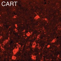
Plasma membrane and vesicular glutamate transporter mRNAs/proteins in hypothalamic neurons that regulate body weight.
Maria Collin, Matilda Bäckberg, Marie-Louise Ovesjö, Gilberto Fisone, Robert H Edwards, Fumino Fujiyama, Björn Meister
The European journal of neuroscience
18
1265-78
2003
Zobrazit abstrakt
After synaptic release, glutamate is taken up by the nerve terminal via a plasma membrane-associated protein termed excitatory amino acid transporter 3 (EAAT3). Following entry into the nerve terminal, glutamate is pumped into synaptic vesicles by a vesicular transport system. Three different vesicular glutamate transporter proteins (VGLUT1-3) representing unique markers for glutamatergic neurons were recently characterized. The presence of EAAT3, glutaminase and VGLUT1-3 was examined in mouse, rat and rabbit species at mRNA and protein levels in hypothalamic neurons which are involved in the regulation of body weight using in situ hybridization and immunohistochemistry. EAAT3 and glutaminase mRNAs were demonstrated in all parts of the arcuate nucleus in the dorsomedial and ventromedial hypothalamic nuclei and lateral hypothalamic area. VGLUT1 mRNA was present in the magnocellular lateral hypothalamic nucleus. VGLUT2 mRNA was demonstrated in a subpopulation of neurons in the arcuate nucleus and in the ventromedial and dorsomedial hypothalamic nuclei and lateral hypothalamic area. Few VGLUT3 mRNA expressing neurons were scattered throughout the medial and lateral hypothalamus. EAAT3-like immunoreactivity (-li) was demonstrated in glutamate, neuropeptide Y (NPY), agouti-related peptide (AGRP), pro-opiomelanocortin (POMC), cocaine and amphetamine-regulated transcript (CART), melanin-concentrating hormone and orexin-immunoreactive (-ir) neurons. VGLUT2-li could only be demonstrated in POMC- and CART-ir neurons of the ventrolateral arcuate nucleus. The results show that key neurons involved in regulation of energy balance are glutamatergic and/or densely innervated by glutamatergic nerve terminals. Whereas orexigenic NPY/AGRP neurons situated in the ventromedial part of the arcuate nucleus are mainly GABAergic, it is shown that several anorexigenic POMC/CART neurons of the ventromedial arcuate nucleus are most likely glutamatergic [corrected]. | 12956725
 |
5-HT1A receptor immunoreactivity in hypothalamic neurons involved in body weight control.
Maria Collin, Matilda Bäckberg, Kristin Onnestam, Björn Meister
Neuroreport
13
945-51
2002
Zobrazit abstrakt
Serotonin (5-hydroxytryptamine; 5-HT) is a regulator of feeding behavior. The effect of serotonin on food intake is believed to be primarily mediated via 5-HT(1A) and 5-HT(2C) receptors, which both are expressed in hypothalamic regions implicated in regulation of feeding behavior. Using an antiserum to the 5-HT(1A) receptor, immunoreactive neurons were observed in the rat supraoptic, paraventricular, arcuate and ventromedial nuclei and lateral hypothalamic area. 5-HT(1A) receptor immunoreactivity was demonstrated in neuropeptide Y-, agouti-related peptide-, proopiomelanocortin- and cocaine- and amphetamine-regulated transcript-containing neurons of the arcuate nucleus. In the lateral hypothalamus, 5-HT(1A) receptor immunoreactivity was observed in melanin-concentrating hormone- and orexin-containing neurons. The results suggest that serotonin via postsynaptic 5-HT(1A) receptors affects the release of peptides regulating food intake. | 12004196
 |
Orexin receptor-1 (OX-R1) immunoreactivity in chemically identified neurons of the hypothalamus: focus on orexin targets involved in control of food and water intake.
Matilda Bäckberg, Guillaume Hervieu, Shelagh Wilson, Björn Meister
The European journal of neuroscience
15
315-28
2002
Zobrazit abstrakt
The neuropeptides orexin-A and orexin-B are produced in neurons of the lateral hypothalamic area and have been implicated to be involved in the regulation of food/water intake and sleep-wake control. The orexins act at two different G-protein-coupled orexin receptors (OX-R1 and OX-R2) that are derived from separate genes and expressed differentially throughout the central nervous system. In the present study, we have used a polyclonal antipeptide antiserum to analyse in detail the distribution of OX-R1-immunoreactive neurons in the rat hypothalamus. In order to identify the chemical mediators of orexin action in the hypothalamus, the OX-R1-containing neurons were characterized with regard to the content of peptides shown previously to affect ingestive and drinking behaviour. Neurons containing OX-R1 immunoreactivity were widely distributed in the hypothalamus with cell bodies located in the suprachiasmatic, periventricular, paraventricular (both magno- and parvocellular division), supraoptic, arcuate, ventromedial, dorsomedial and tuberomammillary nuclei and the lateral hypothalamic area. In magnocellular neurons of the paraventricular and supraoptic nuclei, OX-R1 immunoreactivity was seen in both vasopressin- and oxytocin-containing neurons. OX-R1 immunoreactivity was demonstrated in vasopressin and vasoactive intestinal polypeptide (VIP) neurons of the suprachiasmatic nucleus, in somatostatin neurons of the periventricular nucleus and in corticotropin-releasing hormone (CRH) neurons of the parvocellular paraventricular nucleus. In the arcuate nucleus, OX-R1 immunoreactivity was present in neuropeptide Y (NPY) and agouti-related peptide (AGRP) neurons of the ventromedial part as well as in proopiomelanocortin (POMC) and cocaine- and amphetamine-regulated transcript (CART) neurons of the ventrolateral division. In the lateral hypothalamic area, OX-R1 immunoreactivity was demonstrated in melanin-concentrating hormone (MCH)- and orexin-containing neurons. In the hypothalamic tuberomammillary nucleus, OX-R1-immunoreactivity was shown in many histamine-containing neurons. The results support the idea that orexins have important actions on hypothalamic neurons that control food intake and fluid balance, but also that orexins may regulate other neuroendocrine systems. | 11849298
 |
Control of food intake via leptin receptors in the hypothalamus.
Meister, B
Vitam. Horm., 59: 265-304 (2000)
1999
Zobrazit abstrakt
Food intake is regulated via neural circuits located in the hypothalamus. During the past decade our knowledge on the specific mediators and neuronal networks that regulate food intake and body weight has increased dramatically. An important contribution to the understanding of hypothalamic control of food intake has been the characterization of the ob gene product (leptin) via positional cloning. Absence of circulating, functionally active, leptin hormone results in massive obesity as seen in ob/ob mice. Leptin inhibits food intake and increases energy expenditure via an interaction with specific leptin receptors located in the hypothalamus. Leptin receptors, of which there are several splice variants (Ob-Ra through Ob-Re), belong to the superfamily of cytokine receptors, which use the JAK-STAT pathway of signal transduction. Obese db/db mice, which have a mutation in the db locus, are unable to perform JAK-STAT signal transduction due to absence of functionally active (long form; Ob-Rb) leptin receptors. Ob-Rb is primarily expressed in the hypothalamus, with particularly high levels in the arcuate, paraventricular, and dorsomedial nuclei and in the lateral hypothalamic area. The abundance of leptin receptors in the ventromedial and lateral hypothalamus supports early observations that these two regions are intimately associated with the regulation of food intake. Leptin receptors have been identified in neuropeptide Y (NPY)/lagouti-related peptide (AgRP)- and proopiomelanocortin (POMC)/cocaine- and amphetamine-regulated transcript (CART)-containing neurons of the ventromedial and ventrolateral arcuate nucleus, respectively, and in melanin-concentrating hormone (MCH)- and hypocretin/orexin-containing neurons of the lateral hypothalamus, suggesting that the above-mentioned messengers are mediators of leptin's action in the hypothalamus. Indeed, functional studies show that NPY, AgRP, POMC-derived peptides, CART, MCH, and hypocretins/orexins all are important regulators of food intake. Leptin is essential for normal body weight balance, but the exact mechanisms by which leptin activates hypothalamic neuronal circuitries is known to a limited extent. In order to find pharmaceutical approaches to treat obesity, further studies will be needed to reveal the exact mechanisms by which leptin lowers body weight and which role leptin and leptin receptors have in the pathogenesis of human obesity. | 10714243
 |
Hypothalamic CART is a new anorectic peptide regulated by leptin.
Kristensen, P, et al.
Nature, 393: 72-6 (1998)
1998
Zobrazit abstrakt
The mammalian hypothalamus strongly influences ingestive behaviour through several different signalling molecules and receptor systems. Here we show that CART (cocaine- and amphetamine-regulated transcript), a brain-located peptide, is a satiety factor and is closely associated with the actions of two important regulators of food intake, leptin and neuropeptide Y. Food-deprived animals show a pronounced decrease in expression of CART messenger RNA in the arcuate nucleus. In animal models of obesity with disrupted leptin signalling, CART mRNA is almost absent from the arcuate nucleus. Peripheral administration of leptin to obese mice stimulates CART mRNA expression. When injected intracerebroventricularly into rats, recombinant CART peptide inhibits both normal and starvation-induced feeding, and completely blocks the feeding response induced by neuropeptide Y. An antiserum against CART increases feeding in normal rats, indicating that CART may be an endogenous inhibitor of food intake in normal animals. | 9590691
 |
Characterization of the human cDNA and genomic DNA encoding CART: a cocaine- and amphetamine-regulated transcript.
Douglass, J and Daoud, S
Gene, 169: 241-5 (1996)
1996
Zobrazit abstrakt
PCR differential display screening has recently identified a rat mRNA termed CART (cocaine- and amphetamine-regulated transcript) which is transcriptionally regulated in the striatum following acute administration of psychomotor stimulants. The endogenous CART transcript is expressed in diverse rat brain structures, as well as endocrine tissues. The deduced CART protein contains a hydrophobic signal sequence, suggesting that it may be targeted for secretion. Thus, the CART protein may represent a novel neuroendocrine signaling molecule. The study described here represents a complete analysis of the human CART cDNA and gene. The complete nucleotide (nt) sequence of the approx. 900-nt human CART transcript is contained within three distinct exons, with the entire human CART gene localized to a segment of genomic DNA approx. 2 kb in length. The human CART cDNA sequence is 80% identical to the corresponding rat cDNA, with 92% homology observed within the deduced protein-coding region. Third-nt changes account for most of the latter differences, with CART exhibiting 95% identity between these two species at the amino-acid sequence level. PCR/Southern blot analysis of DNA isolated from human/rodent somatic cell hybrid panels localizes the CART gene to human chromosome 5. Lastly, Northern blot analysis reveals that the gross pattern of distribution of CART mRNA in human brain is similar to that previously observed in rat. These overall similarities suggest that CART plays a conserved role within the mammalian neuroendocrine system. | 8647455
 |
PCR differential display identifies a rat brain mRNA that is transcriptionally regulated by cocaine and amphetamine.
Douglass, J, et al.
J. Neurosci., 15: 2471-81 (1995)
1994
Zobrazit abstrakt
Neuronal plasticity associated with both short- and long-term administration of psychomotor stimulants involves alterations in specific patterns of gene expression. In order to screen for brain region specific mRNAs which are transcriptionally regulated by acute cocaine and amphetamine, PCR differential display was employed. This approach identified a previously uncharacterized mRNA whose relative levels in the striatum are induced four- to fivefold by acute psychomotor stimulant administration. Isolation and characterization of corresponding cDNA clones resulted in complete nucleotide sequence analysis, including prediction of the encoded protein product. Alternate polyA site utilization in the predicted 3' noncoding region results in the appearance of an RNA doublet, approximately 700 and 900 bases in length, following Northern analysis. A presumed alternate splicing event further generates diversity within the transcripts, and results in the presence or absence of an in-frame 39 base insert within the putative protein coding region. As a result, the predicted translation products are either 129 or 116 amino acids in length. A common hydrophobic leader sequence at the amino terminus is present within each predicted polypeptide, suggesting that the protein product is targeted for entry into the secretory pathway. Basal expression of the RNA doublet is limited to neuroendocrine tissues, further implying that the protein product plays a functional role in both neuronal and endocrine tissues. | 7891182
 |


