Our broad portfolio consists of multiplex panels that allow you to choose, within the panel, analytes that best meet your needs. On a separate tab you can choose the premixed cytokine format or a single plex kit.
Cell Signaling Kits & MAPmates™
Choose fixed kits that allow you to explore entire pathways or processes. Or design your own kits by choosing single plex MAPmates™, following the provided guidelines.
The following MAPmates™ should not be plexed together:
-MAPmates™ that require a different assay buffer
-Phospho-specific and total MAPmate™ pairs, e.g. total GSK3β and GSK3β (Ser 9)
-PanTyr and site-specific MAPmates™, e.g. Phospho-EGF Receptor and phospho-STAT1 (Tyr701)
-More than 1 phospho-MAPmate™ for a single target (Akt, STAT3)
-GAPDH and β-Tubulin cannot be plexed with kits or MAPmates™ containing panTyr
.
Catalogue Number
Ordering Description
Qty/Pack
List
This item has been added to favorites.
Select A Species, Panel Type, Kit or Sample Type
To begin designing your MILLIPLEX® MAP kit select a species, a panel type or kit of interest.
Custom Premix Selecting "Custom Premix" option means that all of the beads you have chosen will be premixed in manufacturing before the kit is sent to you.
Catalogue Number
Ordering Description
Qty/Pack
List
This item has been added to favorites.
Species
Panel Type
Selected Kit
Qty
Catalogue Number
Ordering Description
Qty/Pack
List Price
96-Well Plate
Qty
Catalogue Number
Ordering Description
Qty/Pack
List Price
Add Additional Reagents (Buffer and Detection Kit is required for use with MAPmates)
Qty
Catalogue Number
Ordering Description
Qty/Pack
List Price
48-602MAG
Buffer Detection Kit for Magnetic Beads
1 Kit
Space Saver Option Customers purchasing multiple kits may choose to save storage space by eliminating the kit packaging and receiving their multiplex assay components in plastic bags for more compact storage.
This item has been added to favorites.
The Product Has Been Added To Your Cart
You can now customize another kit, choose a premixed kit, check out or close the ordering tool.
Attention: We have moved. Merck Millipore products are no longer available for purchase on MerckMillipore.com.Learn More
PC213L
Sigma-AldrichAnti-GABA Rabbit pAb
Anti-GABA, rabbit polyclonal, recognizes GABA in rat thalamus and cerebellum. It is validated for use in immunohistochemistry on frozen sections.
More>>Anti-GABA, rabbit polyclonal, recognizes GABA in rat thalamus and cerebellum. It is validated for use in immunohistochemistry on frozen sections. Less<<
Anti-GABA Rabbit pAb MSDS (material safety data sheet) or SDS, CoA and CoQ, dossiers, brochures and other available documents.
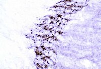
Detection of rat GABA by immunohistochemistry. Sample: Rat thalamus and cerebellum. Primary antibody: Anti-GABA Rabbit pAb (Cat. No. PC213L) (1:20,000). Detection: fluorescence.
Frozen Sections (1:12,000-1:15,000, ABC detection or 1:3000-1:5000, PAP detection; see comments)
Application Comments
Antibody specificity was examined in the rat thalamus and cerebellum. Glutaraldehyde is a necessary component of fixation for use with this antibody. Higher concentrations of glutaraldehyde (1%-2%) may be used as needed. The specificity of the antibody for GABA was evaluated using a competitive inhibition ELISA. A GABA conjugate control completely eliminated labeling, while 1000-fold excess of the following conjugates: alanine, aspartate, beta alanine, glutamate, glycine, taurine, and tyrosine did not inhibit the binding to the GABA conjugate control. Antibody should be titrated for optimal results in individual systems.
Biological Information
Immunogen
GABA
Immunogen
Rat
Host
Rabbit
Isotype
IgG
Species Reactivity
Rat
Antibody Type
Polyclonal Antibody
Physicochemical Information
Dimensions
Materials Information
Toxicological Information
Safety Information according to GHS
Safety Information
Product Usage Statements
Storage and Shipping Information
Ship Code
Ambient Temperature Only
Toxicity
Standard Handling
Storage
-20°C
Do not freeze
Ok to freeze
Special Instructions
Reconstitute the lyophilized antibody with 100 µl sterile distilled H₂O. For storage, dilute with sterile PBS or Tris buffer at dilutions no higher than 1:10. Reconstitute the entire contents of the vial; during shipment and handling portions of the lyophilized pellet may have become dislodged and may not be in the bottom of the vial. Following reconstitution, aliquot and freeze (-20°C). Stock solutions are stable for up to 6 months at -20°C. Avoid freeze/thaw cycles of solutions.
Note that this data sheet is not lot-specific and is representative of the current specifications for this product. Please consult the vial label and the certificate of analysis for information on specific lots. Also note that shipping conditions may differ from storage conditions.
Revision
09-October-2007 RFH
Synonyms
Anti-Gamma Amino-Butyric Acid
Application
Frozen Sections (1:12,000-1:15,000, ABC detection or 1:3000-1:5000, PAP detection; see comments)
Application Data
Detection of rat GABA by immunohistochemistry. Sample: Rat thalamus and cerebellum. Primary antibody: Anti-GABA Rabbit pAb (Cat. No. PC213L) (1:20,000). Detection: fluorescence.
Description
Rabbit polyclonal antibody supplied as lyophilized undiluted serum. Recognizes GABA.
Background
Neurotransmitters can be defined as chemical substances that are released by the presynaptic neuron before being bound by receptors on the postsynaptic cell surface. It has been well established that the nervous system utilizes neurotransmitters for cell signaling. Neurotransmitters can be divided into two groups; low molecular weight transmitter molecules such as biogenic amines, purines, or amino acids, and bioactive peptides, of which more than 50 members have been identified in the mammalian central nervous system. Many low molecular weight neurotransmitters are produced within the axon terminal from common cellular metabolites and packaged into synaptic vesicles. In contrast, neuropeptides are synthesized in the cell body and packaged into large dense core vesicles for delivery to the presynaptic membrane. The transport and delivery of vesicles containing neurotransmitter is a highly regulated and complex process. GABA (g-aminobutyric acid) is the major inhibitory neurotransmitter in the central nervous system that is involved in the pathogenesis of certain neurological and psychiatric disorders. GABA is produced from glutamic acid in a reaction catalyzed by glutamic acid decarboxylase. GABA interacts with the GABAA and GABAB receptors, which are widely distributed throughout the nervous system and in a variety of cell types. The immunolocalization, neuronal distribution, and vesicular compartmentalization of neurotransmitter molecules and their precursors are important for understanding the modulation, regulation, and function of neurotransmitters in the nervous system.
Host
Rabbit
Immunogen species
Rat
Immunogen
GABA
Isotype
IgG
Species
rat
Positive control
Rat thalamus or cerebellum
Form
Lyophilized
Formulation
Undiluted serum.
Preservative
None
Comments
Antibody specificity was examined in the rat thalamus and cerebellum. Glutaraldehyde is a necessary component of fixation for use with this antibody. Higher concentrations of glutaraldehyde (1%-2%) may be used as needed. The specificity of the antibody for GABA was evaluated using a competitive inhibition ELISA. A GABA conjugate control completely eliminated labeling, while 1000-fold excess of the following conjugates: alanine, aspartate, beta alanine, glutamate, glycine, taurine, and tyrosine did not inhibit the binding to the GABA conjugate control. Antibody should be titrated for optimal results in individual systems.
Storage
-20°C
Do Not Freeze
Ok to freeze
Special Instructions
Reconstitute the lyophilized antibody with 100 µl sterile distilled H₂O. For storage, dilute with sterile PBS or Tris buffer at dilutions no higher than 1:10. Reconstitute the entire contents of the vial; during shipment and handling portions of the lyophilized pellet may have become dislodged and may not be in the bottom of the vial. Following reconstitution, aliquot and freeze (-20°C). Stock solutions are stable for up to 6 months at -20°C. Avoid freeze/thaw cycles of solutions.