An accumulation of non-farnesylated prelamin A causes cardiomyopathy but not progeria.
Davies, BS; Barnes, RH; Tu, Y; Ren, S; Andres, DA; Spielmann, HP; Lammerding, J; Wang, Y; Young, SG; Fong, LG
Human molecular genetics
19
2682-94
2010
Show Abstract
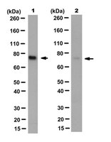
Lamin A is formed from prelamin A by four post-translational processing steps-farnesylation, release of the last three amino acids of the protein, methylation of the farnesylcysteine and the endoproteolytic release of the C-terminal 15 amino acids (including the farnesylcysteine methyl ester). When the final processing step does not occur, a farnesylated and methylated prelamin A accumulates in cells, causing a severe progeroid disease, restrictive dermopathy (RD). Whether RD is caused by the retention of farnesyl lipid on prelamin A, or by the retention of the last 15 amino acids of the protein, is unknown. To address this issue, we created knock-in mice harboring a mutant Lmna allele (LmnanPLAO) that yields exclusively non-farnesylated prelamin A (and no lamin C). These mice had no evidence of progeria but succumbed to cardiomyopathy. We suspected that the non-farnesylated prelamin A in the tissues of these mice would be strikingly mislocalized to the nucleoplasm, but this was not the case; most was at the nuclear rim (indistinguishable from the lamin A in wild-type mice). The cardiomyopathy could not be ascribed to an absence of lamin C because mice expressing an otherwise identical knock-in allele yielding only wild-type prelamin A appeared normal. We conclude that lamin C synthesis is dispensable in mice and that the failure to convert prelamin A to mature lamin A causes cardiomyopathy (at least in the absence of lamin C). The latter finding is potentially relevant to the long-term use of protein farnesyltransferase inhibitors, which lead to an accumulation of non-farnesylated prelamin A. | 20421363
 |
Genetic studies on the functional relevance of the protein prenyltransferases in skin keratinocytes.
Lee, R; Chang, SY; Trinh, H; Tu, Y; White, AC; Davies, BS; Bergo, MO; Fong, LG; Lowry, WE; Young, SG
Human molecular genetics
19
1603-17
2010
Show Abstract
The modification of proteins with farnesyl or geranylgeranyl lipids, a process called protein prenylation, facilitates interactions of proteins with membrane surfaces. Protein prenylation is carried out by a pair of cytosolic enzymes, protein farnesyltransferase (FTase) and protein geranylgeranyltransferase type I (GGTase-I). FTase and GGTase-I have attracted interest as therapeutic targets for both cancer and progeria, but very little information exists on the importance of these enzymes for homeostasis of normal tissues. One study actually suggested that FTase is entirely dispensable. To explore the importance of the protein prenyltransferases for normal tissues, we used conditional knockout alleles for Fntb and Pggt1b (which encode the beta-subunits of FTase and GGTase-I, respectively) and a keratin 14-Cre transgene to create mice lacking FTase or GGTase-I in skin keratinocytes. Keratinocyte-specific Fntb knockout mice were viable but developed severe alopecia. Although hair follicles appeared normal during development, they were morphologically abnormal after birth, and ultrastructural and immunohistochemical studies revealed many apoptotic cells. The interfollicular epidermis of Fntb-deficient mice appeared normal; however, keratinocytes from these mice could not proliferate in culture. As expected, non-farnesylated prelamin A and non-farnesylated DNAJA1 accumulated in Fntb-deficient keratinocytes. Keratinocyte-specific Pggt1b knockout mice survived development but died shortly after birth. Like Fntb-deficient keratinocytes, Pggt1b-deficient keratinocytes did not proliferate in culture. Thus, both FTase and GGTase-I are required for the homeostasis of skin keratinocytes. | 20106865
|