Our broad portfolio consists of multiplex panels that allow you to choose, within the panel, analytes that best meet your needs. On a separate tab you can choose the premixed cytokine format or a single plex kit.
Cell Signaling Kits & MAPmates™
Choose fixed kits that allow you to explore entire pathways or processes. Or design your own kits by choosing single plex MAPmates™, following the provided guidelines.
The following MAPmates™ should not be plexed together:
-MAPmates™ that require a different assay buffer
-Phospho-specific and total MAPmate™ pairs, e.g. total GSK3β and GSK3β (Ser 9)
-PanTyr and site-specific MAPmates™, e.g. Phospho-EGF Receptor and phospho-STAT1 (Tyr701)
-More than 1 phospho-MAPmate™ for a single target (Akt, STAT3)
-GAPDH and β-Tubulin cannot be plexed with kits or MAPmates™ containing panTyr
.
Catalogue Number
Ordering Description
Qty/Pack
List
This item has been added to favorites.
Select A Species, Panel Type, Kit or Sample Type
To begin designing your MILLIPLEX® MAP kit select a species, a panel type or kit of interest.
Custom Premix Selecting "Custom Premix" option means that all of the beads you have chosen will be premixed in manufacturing before the kit is sent to you.
Catalogue Number
Ordering Description
Qty/Pack
List
This item has been added to favorites.
Species
Panel Type
Selected Kit
Qty
Catalogue Number
Ordering Description
Qty/Pack
List Price
96-Well Plate
Qty
Catalogue Number
Ordering Description
Qty/Pack
List Price
Add Additional Reagents (Buffer and Detection Kit is required for use with MAPmates)
Qty
Catalogue Number
Ordering Description
Qty/Pack
List Price
48-602MAG
Buffer Detection Kit for Magnetic Beads
1 Kit
Space Saver Option Customers purchasing multiple kits may choose to save storage space by eliminating the kit packaging and receiving their multiplex assay components in plastic bags for more compact storage.
This item has been added to favorites.
The Product Has Been Added To Your Cart
You can now customize another kit, choose a premixed kit, check out or close the ordering tool.
The quantity field is empty. Please enter a quantity of 1 or more to add items to your cart.
Description
Overview
Recognizes the ~37 kDa PCNA protein in MCF-7 cells.
Catalogue Number
PC474
Brand Family
Calbiochem®
Synonyms
Anti-Proliferating Cell Nuclear Antigen
Application Data
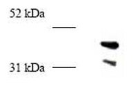
Detection of human PCNA by immunoprecipitation. Sample: Whole cell lysate from MCF7 cells. Antibodyfor IP: Anti-PCNA (Ab-1) Mouse mAb (PC10) (Cat. No. NA03) (1 µg/ml). Anti-PCNA (247-261) (Ab-5) Rabbit pAb (Cat. No. PC474) (1:1000). Detection: chemiluminescence. A nonspecific band appears at ~45 kDa in the sample lysate and in all the controls.
References
References
Miyachi, K., et al. 1990. J. Immunol.121, 2228. Waseem, N.H. and Lane, D.P. 1990. Science96, 121. Suzuka, I., et al. 1989. Proc. Natl. Acad. Sci. USA86, 3189. Bravo, R., et al. 1987. Nature (London)326, 515. Bravo, R. and MacDonald-Bravo, H. 1987. J. Cell. Biol.105, 1549.
Anti-PCNA (247-261) (Ab-5) Rabbit pAb Certificates of Analysis
Title
Lot Number
PC474
References
Reference overview
Miyachi, K., et al. 1990. J. Immunol.121, 2228. Waseem, N.H. and Lane, D.P. 1990. Science96, 121. Suzuka, I., et al. 1989. Proc. Natl. Acad. Sci. USA86, 3189. Bravo, R., et al. 1987. Nature (London)326, 515. Bravo, R. and MacDonald-Bravo, H. 1987. J. Cell. Biol.105, 1549.
Data Sheet
Note that this data sheet is not lot-specific and is representative of the current specifications for this product. Please consult the vial label and the certificate of analysis for information on specific lots. Also note that shipping conditions may differ from storage conditions.
Revision
10-March-2008 JSW
Synonyms
Anti-Proliferating Cell Nuclear Antigen
Application
Immunoprecipitation (1:250-1:1000)
Application Data
Detection of human PCNA by immunoprecipitation. Sample: Whole cell lysate from MCF7 cells. Antibodyfor IP: Anti-PCNA (Ab-1) Mouse mAb (PC10) (Cat. No. NA03) (1 µg/ml). Anti-PCNA (247-261) (Ab-5) Rabbit pAb (Cat. No. PC474) (1:1000). Detection: chemiluminescence. A nonspecific band appears at ~45 kDa in the sample lysate and in all the controls.
Description
Rabbit polyclonal antibody supplied as undiluted serum. Recognizes the ~36-37 kDa PCNA protein.
Background
The proliferating cell nuclear antigen (PCNA) is a ~36-37 kDa protein also known as cyclin. The protein has been identified as a polymerase δ accessory protein and is detected in a cell cycle dependent manner. In early S phase PCNA has a very granular distribution and is absent from the nucleloli. In cells fixed with organic solvents, the PCNA is seen to be strongly associated in the nuclear regions where DNA synthesis is occurring, whereas in cell fixed with aldehydes the staining is more diffuse but intense and occurs throughout the cell cycle. This is due to the presence of two basic forms of the PCNA protein: a soluble form which is sensitive to organic fixation and not involved in replication and a second form which is insoluble and associated with on going DNA synthesis. PCNA is a very conserved protein present not only in mammals but also in plant cells. Patients suffering from SLE have been shown to have autoantibodies against PCNA.
Host
Rabbit
Immunogen species
Human
Immunogen
a synthetic peptide corresponding to amino acids 247-261 of human PCNA
Isotype
IgG
Species
human
Positive control
MCF7 cells
Form
Liquid
Formulation
Undiluted serum.
Preservative
≤0.1% sodium azide
Comments
Will immunoprecipitate PCNA when complexed with other proteins. Antibody should be titrated for optimal results in individual systems.
Storage
Avoid freeze/thaw
-20°C
Do Not Freeze
Ok to freeze
Special Instructions
Following initial thaw, aliquot and freeze (-20°C).
Toxicity
Standard Handling
References
Miyachi, K., et al. 1990. J. Immunol.121, 2228. Waseem, N.H. and Lane, D.P. 1990. Science96, 121. Suzuka, I., et al. 1989. Proc. Natl. Acad. Sci. USA86, 3189. Bravo, R., et al. 1987. Nature (London)326, 515. Bravo, R. and MacDonald-Bravo, H. 1987. J. Cell. Biol.105, 1549.