Millipore Sigma Vibrant Logo

NE1017 Anti-Pan-Neuronal Neurofilament Marker Mouse mAb (SMI-311)

View Products on Sigmaaldrich.com
NE1017
  
Retrieving price...
Price could not be retrieved
Minimum Quantity is a multiple of
Maximum Quantity is
Upon Order Completion More Information
You Saved ()
 
Request Pricing
Limited Availability
In Stock 
Discontinued
Limited Quantities Available
Availability to be confirmed
    Remaining : Will advise
      Remaining : Will advise
      Will advise
      Contact Customer Service
      Contact Customer Service


      Contact Customer Service

      Overview

      Replacement Information

      Key Spec Table

      Species ReactivityHostAntibody Type
      MaMMonoclonal Antibody
      Description
      Overview

      This product has been discontinued.


      Recognizes ~180-200 kDa pan-neuronal neurofilament marker in rat central nervous system cytoskeletal preparations.

      Catalogue NumberNE1017
      Brand Family Calbiochem®
      Application Data
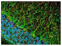
      Detection of rat pan-neuronal neurofilament marker by staining frozen sections. Sample: Rat brain. Primary antibody: Anti-Pan-Neuronal Neurofilament Marker Mouse mAb (SMI-311) (Cat. No. NE1017) (1:1000). Detection: fluorescence (green) with Hoechst 33342 counterstain.
      References
      ReferencesAgostino, A., et al. 2003. Hum. Mol. Genet. 12, 399.
      Tsunoda, I., et al. 2003. Am. J. Pathol. 162, 1259.
      Ulfig, N., et al. 1998. Cell Tissue Res. 291, 433.
      Product Information
      FormLiquid
      FormulationUndiluted ascites.
      Positive controlRat brain or rat CNS cytoskeletal preparations
      Preservative≤0.1% sodium azide
      Quality LevelMQ100
      Applications
      Key Applications Enzyme-Linked Immunosorbent Assay
      Frozen Sections
      Immunoblotting (Western Blotting)
      Immunocytochemistry
      Paraffin Sections
      Application NotesELISA (1:1000)
      Frozen Sections (1:1000, see comments)
      Immunoblotting (1:1000)
      Immunocytochemistry (1:1000, see comments)
      Paraffin Sections (1:1000, heat pre-treatment required, see comments)
      Application CommentsThis antibody cocktail was selected to provide a specific marker for neurons in tissue sections and cultured cells. In contrast to individual anti-nonphosphoneurofilament antibodies that identify different subsets of neurons, this cocktail is a convenient marker for neurons in general and their differentiation from non-neuronal cells. Also useful as an early marker of neuronal migration and differentiation in human fetal development, yielding Golgi-like images without the disadvantages of the lack of selectivity and poor specificity of the Golgi technique. Can be used to trace the "inside-out gradient" of neuron production and differentiation in specifically delineating cell bodies and dendrites. Certain pathologic conditions, such as malnutrition, affect the SMI-311-visualized soma size and dendritic arborization. Tissues and cultured cells can be fixed in a variety of paraformaldehyde- or formaldehyde-containing fixatives, including Bouin's fixative. This antibody does not react well with tissues or cells fixed in glutaraldehyde/paraformaldehyde. For staining paraffin sections it is recommended that de-paraffinized tissues be autoclaved in dH2O for 10 min to expose the epitope. Trypsin pre-treatment will abolish reactivity. Post-fixation in cold methanol or methanol/hydrogen peroxide facilitates access of the antibody to the neurons in frozen sections and thick tissues sections that have been fixed in 4% paraformaldehyde or cultured cells. For tissues that have not been paraffin-embedded and have been stored for long periods of time in formaldehyde fixatives, it is recommended that the tissues (up to 0.5 cm thick) be boiled in Tris-buffered saline, pH 9.0 for 15 min prior to sectioning. Antibody should be titrated for optimal results in individual systems.
      Biological Information
      Immunogenhomogenized hypothalami extracted from Fischer 344 rat brain
      ImmunogenRat
      CloneSMI-311
      HostMouse
      IsotypeIgG₁, IgM cocktail
      Species Reactivity
      • Mammals
      Antibody TypeMonoclonal Antibody
      Physicochemical Information
      Dimensions
      Materials Information
      Toxicological Information
      Safety Information according to GHS
      Safety Information
      Product Usage Statements
      Storage and Shipping Information
      Ship Code Dry Ice Only
      Toxicity Standard Handling
      Storage -20°C
      Avoid freeze/thaw Avoid freeze/thaw
      Do not freeze Ok to freeze
      Special InstructionsFollowing initial thaw, aliquot and freeze (-20°C).
      Packaging Information
      Transport Information
      Supplemental Information
      Specifications
      Global Trade Item Number
      Catalogue Number GTIN
      NE1017 0

      Documentation

      Anti-Pan-Neuronal Neurofilament Marker Mouse mAb (SMI-311) MSDS

      Title

      Safety Data Sheet (SDS) 

      Anti-Pan-Neuronal Neurofilament Marker Mouse mAb (SMI-311) Certificates of Analysis

      TitleLot Number
      NE1017

      References

      Reference overview
      Agostino, A., et al. 2003. Hum. Mol. Genet. 12, 399.
      Tsunoda, I., et al. 2003. Am. J. Pathol. 162, 1259.
      Ulfig, N., et al. 1998. Cell Tissue Res. 291, 433.
      Data Sheet

      Note that this data sheet is not lot-specific and is representative of the current specifications for this product. Please consult the vial label and the certificate of analysis for information on specific lots. Also note that shipping conditions may differ from storage conditions.

      Revision01-October-2007 RFH
      ApplicationELISA (1:1000)
      Frozen Sections (1:1000, see comments)
      Immunoblotting (1:1000)
      Immunocytochemistry (1:1000, see comments)
      Paraffin Sections (1:1000, heat pre-treatment required, see comments)
      Application Data
      Detection of rat pan-neuronal neurofilament marker by staining frozen sections. Sample: Rat brain. Primary antibody: Anti-Pan-Neuronal Neurofilament Marker Mouse mAb (SMI-311) (Cat. No. NE1017) (1:1000). Detection: fluorescence (green) with Hoechst 33342 counterstain.
      DescriptionMouse monoclonal antibody supplied as undiluted ascites. Recognizes the ~180-200 kDa pan-neuronal neurofilament marker protein.
      BackgroundPan-neuronal neurofilament marker is a specific marker for neurons.
      HostMouse
      Immunogen speciesRat
      Immunogenhomogenized hypothalami extracted from Fischer 344 rat brain
      CloneSMI-311
      IsotypeIgG₁, IgM cocktail
      Speciesmammalian
      Positive controlRat brain or rat CNS cytoskeletal preparations
      FormLiquid
      FormulationUndiluted ascites.
      Preservative≤0.1% sodium azide
      CommentsThis antibody cocktail was selected to provide a specific marker for neurons in tissue sections and cultured cells. In contrast to individual anti-nonphosphoneurofilament antibodies that identify different subsets of neurons, this cocktail is a convenient marker for neurons in general and their differentiation from non-neuronal cells. Also useful as an early marker of neuronal migration and differentiation in human fetal development, yielding Golgi-like images without the disadvantages of the lack of selectivity and poor specificity of the Golgi technique. Can be used to trace the "inside-out gradient" of neuron production and differentiation in specifically delineating cell bodies and dendrites. Certain pathologic conditions, such as malnutrition, affect the SMI-311-visualized soma size and dendritic arborization. Tissues and cultured cells can be fixed in a variety of paraformaldehyde- or formaldehyde-containing fixatives, including Bouin's fixative. This antibody does not react well with tissues or cells fixed in glutaraldehyde/paraformaldehyde. For staining paraffin sections it is recommended that de-paraffinized tissues be autoclaved in dH2O for 10 min to expose the epitope. Trypsin pre-treatment will abolish reactivity. Post-fixation in cold methanol or methanol/hydrogen peroxide facilitates access of the antibody to the neurons in frozen sections and thick tissues sections that have been fixed in 4% paraformaldehyde or cultured cells. For tissues that have not been paraffin-embedded and have been stored for long periods of time in formaldehyde fixatives, it is recommended that the tissues (up to 0.5 cm thick) be boiled in Tris-buffered saline, pH 9.0 for 15 min prior to sectioning. Antibody should be titrated for optimal results in individual systems.
      Storage Avoid freeze/thaw
      -20°C
      Do Not Freeze Ok to freeze
      Special InstructionsFollowing initial thaw, aliquot and freeze (-20°C).
      Toxicity Standard Handling
      ReferencesAgostino, A., et al. 2003. Hum. Mol. Genet. 12, 399.
      Tsunoda, I., et al. 2003. Am. J. Pathol. 162, 1259.
      Ulfig, N., et al. 1998. Cell Tissue Res. 291, 433.