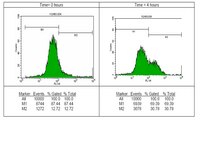
Inactivation of Rb and E2f8 synergizes to trigger stressed DNA replication during erythroid terminal differentiation.
Ghazaryan, S; Sy, C; Hu, T; An, X; Mohandas, N; Fu, H; Aladjem, MI; Chang, VT; Opavsky, R; Wu, L
Molecular and cellular biology
34
2833-47
2014
Pokaż streszczenie
Rb is critical for promoting cell cycle exit in cells undergoing terminal differentiation. Here we show that during erythroid terminal differentiation, Rb plays a previously unappreciated and unorthodox role in promoting DNA replication and cell cycle progression. Specifically, inactivation of Rb in erythroid cells led to stressed DNA replication, increased DNA damage, and impaired cell cycle progression, culminating in defective terminal differentiation and anemia. Importantly, all of these defects associated with Rb loss were exacerbated by the concomitant inactivation of E2f8. Gene expression profiling and chromatin immunoprecipitation (ChIP) revealed that Rb and E2F8 cosuppressed a large array of E2F target genes that are critical for DNA replication and cell cycle progression. Remarkably, inactivation of E2f2 rescued the erythropoietic defects resulting from Rb and E2f8 deficiencies. Interestingly, real-time quantitative PCR (qPCR) on E2F2 ChIPs indicated that inactivation of Rb and E2f8 synergizes to increase E2F2 binding to its target gene promoters. Taken together, we propose that Rb and E2F8 collaborate to promote DNA replication and erythroid terminal differentiation by preventing E2F2-mediated aberrant transcriptional activation through the ability of Rb to bind and sequester E2F2 and the ability of E2F8 to compete with E2F2 for E2f-binding sites on target gene promoters. | Fluorescence Activated Cell Sorting (FACS) | | 24865965
 |
MEIOB exhibits single-stranded DNA-binding and exonuclease activities and is essential for meiotic recombination.
Luo, M; Yang, F; Leu, NA; Landaiche, J; Handel, MA; Benavente, R; La Salle, S; Wang, PJ
Nature communications
4
2788
2013
Pokaż streszczenie
Meiotic recombination enables the reciprocal exchange of genetic material between parental homologous chromosomes, and ensures faithful chromosome segregation during meiosis in sexually reproducing organisms. This process relies on the complex interaction of DNA repair factors and many steps remain poorly understood in mammals. Here we report the identification of MEIOB, a meiosis-specific protein, in a proteomics screen for novel meiotic chromatin-associated proteins in mice. MEIOB contains an OB domain with homology to one of the RPA1 OB folds. MEIOB binds to single-stranded DNA and exhibits 3'-5' exonuclease activity. MEIOB forms a complex with RPA and with SPATA22, and these three proteins co-localize in foci that are associated with meiotic chromosomes. Strikingly, chromatin localization and stability of MEIOB depends on SPATA22 and vice versa. Meiob-null mouse mutants exhibit a failure in meiosis and sterility in both sexes. Our results suggest that MEIOB is required for meiotic recombination and chromosomal synapsis. | | | 24240703
 |
Lower phosphorylation of p38 MAPK blocks the oxidative stress-induced senescence in myeloid leukemic CD34(+)CD38 (-) cells.
Yin Xiao,Ping Zou,Jie Wang,Hui Song,Jing Zou,Lingbo Liu
Journal of Huazhong University of Science and Technology. Medical sciences = Hua zhong ke ji da xue xue bao. Yi xue Ying De wen ban = Huazhong keji daxue xuebao. Yixue Yingdewen ban
32
2011
Pokaż streszczenie
Leukemia seems to depend on a small population of leukemia stem cells (LSCs) for its growth and metastasis. However, the precise surviving mechanisms of LSCs remain obscure. Cellular senescence is an important obstacle for production and surviving of tumor cells. In this study we investigated the activated state of a pathway, in which reactive oxygen species (ROS) induces cellular senescence through DNA damage and phophorylation of p38 MAPK (p38), in myeloid leukemic CD34(+)CD38(-) cells. Bone marrow samples were obtained from patients with acute myeloid leukemia (AML, n=11) and chronic myeloid leukemia (CML, n=9). CD34(+)CD38(-) cells were isolated from mononuclear cells from these bone marrow samples, and K562 and KG1a cells (two kinds of myeloid leukemia cell lines) by mini-magnetic activated cell sorting. Hematopoietic stem cells (HSCs) from human cord blood served as controls. Intracellular ROS level was detected by flow cytometry. DNA damage defined as the γH2AX level was measured by immunofluorescence staining. Real-time RT-PCR was used to detect the expression of p21, a senescence-associated gene. Western blotting and immunofluorescence staining were employed to determine the p38 expression and activation. The proliferation and apoptosis of CD34(+)CD38(-) cells were detected by MTT assay and flow cytometry. Our results showed that ROS and DNA damage were substantially accumulated and p38 was less phosphorated in myeloid leukemic CD34(+)CD38(-) cells as compared with HSCs and H(2)O(2)-induced senescent HSCs. Furthermore, over-phosphorylation of p38 by anisomycin, a selective activator of p38, induced both the senescence-like growth arrest and apoptosis of CD34(+)CD38(-) cells from K562 and KG1a cell lines. These findings suggested that, although excessive accumulation of oxidative DNA damage was present in LSCs, the relatively decreased phosphorylation of p38 might help leukemic cells escape senescence and apoptosis. | | | 22684553
 |
Role of transcriptional corepressor CtBP1 in prostate cancer progression.
Wang, R; Asangani, IA; Chakravarthi, BV; Ateeq, B; Lonigro, RJ; Cao, Q; Mani, RS; Camacho, DF; McGregor, N; Schumann, TE; Jing, X; Menawat, R; Tomlins, SA; Zheng, H; Otte, AP; Mehra, R; Siddiqui, J; Dhanasekaran, SM; Nyati, MK; Pienta, KJ; Palanisamy, N; Kunju, LP; Rubin, MA; Chinnaiyan, AM; Varambally, S
Neoplasia (New York, N.Y.)
14
905-14
2011
Pokaż streszczenie
Transcriptional repressors and corepressors play a critical role in cellular homeostasis and are frequently altered in cancer. C-terminal binding protein 1 (CtBP1), a transcriptional corepressor that regulates the expression of tumor suppressors and genes involved in cell death, is known to play a role in multiple cancers. In this study, we observed the overexpression and mislocalization of CtBP1 in metastatic prostate cancer and demonstrated the functional significance of CtBP1 in prostate cancer progression. Transient and stable knockdown of CtBP1 in prostate cancer cells inhibited their proliferation and invasion. Expression profiling studies of prostate cancer cell lines revealed that multiple tumor suppressor genes are repressed by CtBP1. Furthermore, our studies indicate a role for CtBP1 in conferring radiation resistance to prostate cancer cell lines. In vivo studies using chicken chorioallantoic membrane assay, xenograft studies, and murine metastasis models suggested a role for CtBP1 in prostate tumor growth and metastasis. Taken together, our studies demonstrated that dysregulated expression of CtBP1 plays an important role in prostate cancer progression and may serve as a viable therapeutic target. | | | 23097625
 |
Oncogenic stress sensitizes murine cancers to hypomorphic suppression of ATR.
Schoppy, DW; Ragland, RL; Gilad, O; Shastri, N; Peters, AA; Murga, M; Fernandez-Capetillo, O; Diehl, JA; Brown, EJ
The Journal of clinical investigation
122
241-52
2011
Pokaż streszczenie
Oncogenic Ras and p53 loss-of-function mutations are common in many advanced sporadic malignancies and together predict a limited responsiveness to conventional chemotherapy. Notably, studies in cultured cells have indicated that each of these genetic alterations creates a selective sensitivity to ataxia telangiectasia and Rad3-related (ATR) pathway inhibition. Here, we describe a genetic system to conditionally reduce ATR expression to 10% of normal levels in adult mice to compare the impact of this suppression on normal tissues and cancers in vivo. Hypomorphic suppression of ATR minimally affected normal bone marrow and intestinal homeostasis, indicating that this level of ATR expression was sufficient for highly proliferative adult tissues. In contrast, hypomorphic ATR reduction potently inhibited the growth of both p53-deficient fibrosarcomas expressing H-rasG12V and acute myeloid leukemias (AMLs) driven by MLL-ENL and N-rasG12D. Notably, DNA damage increased in a greater-than-additive fashion upon combining ATR suppression with oncogenic stress (H-rasG12V, K-rasG12D, or c-Myc overexpression), indicating that this cooperative genome-destabilizing interaction may contribute to tumor selectivity in vivo. This toxic interaction between ATR suppression and oncogenic stress occurred without regard to p53 status. These studies define a level of ATR pathway inhibition in which the growth of malignancies harboring oncogenic mutations can be suppressed with minimal impact on normal tissue homeostasis, highlighting ATR inhibition as a promising therapeutic strategy. | Immunofluorescence | Mouse | 22133876
 |
Validation of a flow cytometry based G(2)M delay cell cycle assay for use in evaluating the pharmacodynamic response to Aurora A inhibition.
Estevam J, Danaee H, Liu R, Ecsedy J, Trepicchio WL, Wyant T
J Immunol Methods
2009
Pokaż streszczenie
Pharmacodynamic assays are important aspects for understanding molecularly targeted anticancer agents to investigate the relationship between drug concentration (pharmacokinetics) and drug "effect" or biological activity. As new drug entities are developed that affect DNA cell cycle, a pharmacodynamic assay which measures cell cycle perturbation would be a valuable clinical trial tool. During recent years, flow cytometry has established itself as a useful method to determine the relative nuclear DNA content and percentage of cycling cells of biological specimens. However to date, the analytical validation of cytometry based assays is limited and there is no suitable guidance for method validation of flow cytometry based cell cycle assays. Here we report the validation of a flow cytometry based cell cycle G(2)/M delay assay for use in evaluating the effect of investigational drug MLN8237, a small molecule inhibitor of a mitotic kinase Aurora A, for clinical trial use. The assay method was validated by examining assay robustness, repeatability, reproducibility, precision, and determining the cutoff for a true drug effect based on biostatistical analysis models. Experimental results show that the intra-assay repeatability was less than 20% with an intra-donor variability of less than 40%. The robustness of the assay was less than 30%. Since this is an ex-vivo stimulation assay, variability parameters were expected to be higher. Based on biostatistical modeling, an absolute change in %G(2)M of 5.2% (95% CI) was needed in order to detect a true drug effect. Overall, the assay demonstrated acceptable variability to warrant further in vivo testing. | | | 20887727
 |
Widespread phosphorylation of histone H2AX by species C adenovirus infection requires viral DNA replication.
Nichols, GJ; Schaack, J; Ornelles, DA
Journal of virology
83
5987-98
2009
Pokaż streszczenie
Adenovirus infection activates cellular DNA damage response and repair pathways. Viral proteins that are synthesized before viral DNA replication prevent recognition of viral genomes as a substrate for DNA repair by targeting members of the sensor complex composed of Mre11/Rad50/NBS1 for degradation and relocalization, as well as targeting the effector protein DNA ligase IV. Despite inactivation of these cellular sensor and effector proteins, infection results in high levels of histone 2AX phosphorylation, or gammaH2AX. Although phosphorylated H2AX is a characteristic marker of double-stranded DNA breaks, this modification was widely distributed throughout the nucleus of infected cells and was coincident with the bulk of cellular DNA. H2AX phosphorylation occurred after the onset of viral DNA replication and after the degradation of Mre11. Experiments with inhibitors of the serine-threonine kinases ataxia telangiectasia mutated (ATM), AT- and Rad3-related (ATR), and DNA protein kinase (DNA-PK), the kinases responsible for H2AX phosphorylation, indicate that H2AX may be phosphorylated by ATR during a wild-type adenovirus infection, with some contribution from ATM and DNA-PK. Viral DNA replication appears to be the stimulus for this phosphorylation event, since infection with a nonreplicating virus did not elicit phosphorylation of H2AX. Infected cells also responded to high levels of input viral DNA by localized phosphorylation of H2AX. These results are consistent with a model in which adenovirus-infected cells sense and respond to both incoming viral DNA and viral DNA replication. Pełny tekst artykułu | | | 19321613
 |
Gene deregulation and spatial genome reorganization near breakpoints prior to formation of translocations in anaplastic large cell lymphoma.
Mathas, S; Kreher, S; Meaburn, KJ; Jöhrens, K; Lamprecht, B; Assaf, C; Sterry, W; Kadin, ME; Daibata, M; Joos, S; Hummel, M; Stein, H; Janz, M; Anagnostopoulos, I; Schrock, E; Misteli, T; Dörken, B
Proceedings of the National Academy of Sciences of the United States of America
106
5831-6
2009
Pokaż streszczenie
Although the identification and characterization of translocations have rapidly increased, little is known about the mechanisms of how translocations occur in vivo. We used anaplastic large cell lymphoma (ALCL) with and without the characteristic t(2;5)(p23;q35) translocation to study the mechanisms of formation of translocations and of ALCL transformation. We report deregulation of several genes located near the ALCL translocation breakpoint, regardless of whether the tumor contains the t(2;5). The affected genes include the oncogenic transcription factor Fra2 (located on 2p23), the HLH protein Id2 (2p25), and the oncogenic tyrosine kinase CSF1-receptor (5q33.1). Their up-regulation promotes cell survival and repression of T cell-specific gene expression programs that are characteristic for ALCL. The deregulated genes are in spatial proximity within the nuclear space of t(2;5)-negative ALCL cells, facilitating their translocation on induction of double-strand breaks. These data suggest that deregulation of breakpoint-proximal genes occurs before the formation of translocations, and that aberrant transcriptional activity of genomic regions is linked to their propensity to undergo chromosomal translocations. Also, our data demonstrate that deregulation of breakpoint-proximal genes has a key role in ALCL. | | | 19321746
 |
An optimized method for measurement of gamma-H2AX in blood mononuclear and cultured cells.
Aida Muslimovic,Ismail Hassan Ismail,Yue Gao,Ola Hammarsten
Nature protocols
3
2008
Pokaż streszczenie
Phosphorylation of histone protein H2AX on serine 139 (gamma-H2AX) occurs at sites flanking DNA double-stranded breaks (DSBs) and can provide a measure of the number of DSBs within a cell. We describe a flow cytometry-based method optimized to measure gamma-H2AX in nonfixed mononuclear blood cells as well as in cultured cells, which is more sensitive and involves less steps compared with protocols involving fixed cells. This method can be used to monitor induction of gamma-H2AX in mononuclear cells from cancer patients undergoing radiotherapy and for detection of gamma-H2AX throughout the cell cycle in cultured cells. The method is based on the fact that H2AX like other histone proteins are retained in the nucleus when cells are lysed at physiological salt concentrations. Cells are therefore added without fixation to a solution containing detergent to lyse the cells along with a fluorescein isothiocyanate-labeled monoclonal gamma-H2AX antibody, DNA staining dye and blocking agents. The stained nuclei can be analyzed by flow cytometry to monitor the level of gamma-H2AX to determine the level of DSBs and DNA content and to determine the cell cycle stage. The omission of fixation simplifies staining and enhances the sensitivity. This protocol can be completed within 4-6 h. | | | 18600224
 |
Protective role of Puralpha to cisplatin.
Kaminski, R; Darbinyan, A; Merabova, N; Deshmane, SL; White, MK; Khalili, K
Cancer biology & therapy
7
1926-35
2008
Pokaż streszczenie
The nucleic acid-binding protein Puralpha is involved at stalled DNA replication forks, in double-strand break (DSB) DNA repair and the cellular response to DNA replication stress. Puralpha also regulates homologous recombination-directed DNA repair (HRR).Cells lacking Puralpha showed enhanced sensitivity to cisplatin as evaluated by assays for cell viability and cell clonogenicity. This was seen both in Puralpha-negative MEFs and in human glioblastoma cells treated with siRNA directed against Puralpha. MEFs lacking Puralpha also showed enhanced H2AX phosphorylation in response to cisplatin. Repair of a reporter plasmid that had been treated with cisplatin was decreased in a reactivation assay using Puralpha-negative MEFs and the capacity of nuclear extracts from Puralpha-negative MEFs to perform non-homologous end-joining in vitro was also impaired.We investigated the effects of the DNA damage-inducing cancer chemotherapeutic agent cisplatin on mouse embryo fibroblasts (MEFs) from PURA(-/-) knockout mice that lack Puralpha.Puralpha has a role in the cellular response to cisplatin-induced DNA damage and may provide new therapeutic modalities for cisplatin-resistant tumors. | | | 18927497
 |


