Millipore Sigma Vibrant Logo
Attention: We have moved. Merck Millipore products are no longer available for purchase on MerckMillipore.com.Learn More

577801 Anti-Tau Mouse mAb (TAU-5)

577801
Purchase on Sigma-Aldrich

Przegląd

Replacement Information

Tabela kluczowych gatunków

Species ReactivityHostAntibody Type
H, M, R, ShMMonoclonal Antibody

Products

Numer katalogowyOpakowanie Ilość/opak.
577801-100UG Ampulka plastikowa 100 μg
Description
OverviewRecognizes the ~45-68 kDa Tau protein.
Catalogue Number577801
Brand Family Calbiochem®
Application Data
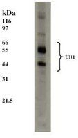
Detection of human tau by immunoblotting. Samples: Whole cell lysate from SH-SY5Y neuronal cells. Primary antibody: Anti-Tau Mouse mAb (TAU-5) (Cat No. 577801) (1:1000). Detection: chemiluminescence.
References
ReferencesPapasozomenos, S.C. and Shanavas, A., 2002. Proc. Natl. Acad. Sci. USA 99, 1140.
Rapoport, M., eta al. 2002. Proc. Natl. Acad. Sci. USA 99, 6364.
Product Information
FormLiquid
FormulationIn PBS.
PreservativeNone
Quality LevelMQ100
Applications
Key Applications Frozen Sections
Immunoblotting (Western Blotting)
Immunoprecipitation
Paraffin Sections
Application NotesFrozen Sections (1-2 µg/ml)
Immunoblotting (1 µg/ml)
Immunoprecipitation (10 µg per 200-500 µg cell lysate)
Paraffin Sections (1-2 µg/ml, heat pre-treatment required)
Application CommentsWhen used for formalin/paraffin embedded sections, staining is enhanced by boiling tissue sections in 10 mM citrate buffer, pH 6.0, for 10-20 min followed by cooling at room temperature for 20 min prior to antibody incubation. Alternate splicing of tau mRNA and differential phosphorylation contribute to the heterogeneity of tau. Does not cross-react with other MAPS of tubulin. Recognizes both native and phosphorylated forms of tau. Variables associated with assay conditions will dictate proper working dilution.
Biological Information
Immunogenpurified bovine microtubule-associated proteins
ImmunogenBovine
Epitopewithin the central region
CloneTAU-5
HostMouse
IsotypeIgG₁
Species Reactivity
  • Human
  • Mouse
  • Rat
  • Sheep
Antibody TypeMonoclonal Antibody
Concentration Label Please refer to vial label for lot-specific concentration
Physicochemical Information
Dimensions
Materials Information
Toxicological Information
Safety Information according to GHS
Safety Information
Product Usage Statements
Storage and Shipping Information
Ship Code Dry Ice Only
Toxicity Standard Handling
Storage ≤ -70°C
Avoid freeze/thaw Avoid freeze/thaw
Do not freeze Ok to freeze
Special InstructionsFollowing initial thaw, aliquot and freeze (-70°C). Do not store diluted antibody without carrier protein if the concentration is <50 µg/ml.
Packaging Information
Transport Information
Supplemental Information
Specifications
Global Trade Item Number
Numer katalogowy GTIN
577801-100UG 04055977265910

Documentation

Anti-Tau Mouse mAb (TAU-5) MSDS

Title

Safety Data Sheet (SDS) 

Anti-Tau Mouse mAb (TAU-5) Certificates of Analysis

TitleLot Number
577801

References

Przegląd literatury
Papasozomenos, S.C. and Shanavas, A., 2002. Proc. Natl. Acad. Sci. USA 99, 1140.
Rapoport, M., eta al. 2002. Proc. Natl. Acad. Sci. USA 99, 6364.

Citations

Tytuł
  • Hui, L., et al. 2012. Life Sci. 91, 1159.
  • Hui, L., et al. 2012. ASN Neuro. 4, 243.
  • Chen, X., et al. 2013. Neurobiol Aging 34, 2370.
  • .
    Data Sheet

    Note that this data sheet is not lot-specific and is representative of the current specifications for this product. Please consult the vial label and the certificate of analysis for information on specific lots. Also note that shipping conditions may differ from storage conditions.

    Revision03-May-2010 JSW
    ApplicationFrozen Sections (1-2 µg/ml)
    Immunoblotting (1 µg/ml)
    Immunoprecipitation (10 µg per 200-500 µg cell lysate)
    Paraffin Sections (1-2 µg/ml, heat pre-treatment required)
    Application Data
    Detection of human tau by immunoblotting. Samples: Whole cell lysate from SH-SY5Y neuronal cells. Primary antibody: Anti-Tau Mouse mAb (TAU-5) (Cat No. 577801) (1:1000). Detection: chemiluminescence.
    DescriptionProtein G purified mouse monoclonal antibody. Recognizes the ~45-68 kDa phosphorylated and unphosphorylated forms of tau.
    BackgroundTau is a neuronal microtubule-associated protein found predominantly in axons. Tau promotes tubulin polymerization and stabilizes microtubules, and it links certain signaling pathways to the cytoskeleton. Hyperphosphorylated tau is the major component of paired helical filaments, the building block of neurofibrillary lesions in Alzheimer’s disease. Hyperphosphorylation impairs the microtubule-binding function of tau, resulting in the destabilization of microtubules, and ultimately leading to degeneration of the affected neurons.
    HostMouse
    Immunogen speciesBovine
    Immunogenpurified bovine microtubule-associated proteins
    Epitopewithin the central region
    CloneTAU-5
    IsotypeIgG₁
    Specieshuman, mouse, rat, sheep
    FormLiquid
    FormulationIn PBS.
    Concentration Label Please refer to vial label for lot-specific concentration
    PreservativeNone
    CommentsWhen used for formalin/paraffin embedded sections, staining is enhanced by boiling tissue sections in 10 mM citrate buffer, pH 6.0, for 10-20 min followed by cooling at room temperature for 20 min prior to antibody incubation. Alternate splicing of tau mRNA and differential phosphorylation contribute to the heterogeneity of tau. Does not cross-react with other MAPS of tubulin. Recognizes both native and phosphorylated forms of tau. Variables associated with assay conditions will dictate proper working dilution.
    Storage Avoid freeze/thaw
    ≤ -70°C
    Do Not Freeze Ok to freeze
    Special InstructionsFollowing initial thaw, aliquot and freeze (-70°C). Do not store diluted antibody without carrier protein if the concentration is <50 µg/ml.
    Toxicity Standard Handling
    ReferencesPapasozomenos, S.C. and Shanavas, A., 2002. Proc. Natl. Acad. Sci. USA 99, 1140.
    Rapoport, M., eta al. 2002. Proc. Natl. Acad. Sci. USA 99, 6364.
    Citation
  • Hui, L., et al. 2012. Life Sci. 91, 1159.
  • Hui, L., et al. 2012. ASN Neuro. 4, 243.
  • Chen, X., et al. 2013. Neurobiol Aging 34, 2370.
  • .