Millipore Sigma Vibrant Logo
Attention: We have moved. Merck Millipore products are no longer available for purchase on MerckMillipore.com.Learn More

NE1013 Anti-PGP9.5 (176-191) Rabbit pAb

NE1013
Purchase on Sigma-Aldrich

Przegląd

Replacement Information

Tabela kluczowych gatunków

Species ReactivityHostAntibody Type
H, M, Porcine, RRbPolyclonal Antibody

Products

Numer katalogowyOpakowanie Ilość/opak.
NE1013-50UL Ampulka plastikowa 50 ul
Description
OverviewRecognizes the ~26 kDa PGP9.5 protein in mouse dorsal root nerve fibers.
Catalogue NumberNE1013
Brand Family Calbiochem®
SynonymsAnti-Neuron Cytoplasmic Protein 9.5, Anti-Ubiquitin Thiolesterase L1, Anti-UCHL1
Application Data
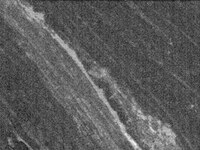
Detection of mouse PGP9.5 by staining frozen sections. Samples: Dorsal root ganglia nerve fibers fixed in paraformaldehyde. Primary antibody: Anti-PGP9.5 (176-191) Rabbit pAb (Cat. No. NE1013) (1:2000) Detection: fluorescence.
References
ReferencesCaballero, O.L., et al. 2002. Oncogene 21, 3003.
Tezel, E., et al. 2000. Clin. Cancer Res. 6, 4764.
Product Information
FormLiquid
FormulationUndiluted serum.
Positive controlMouse dorsal root nerve fibers
Preservative≤0.1% sodium azide
Quality LevelMQ100
Applications
Key Applications Frozen Sections
Immunoblotting (Western Blotting)
Application NotesImmunoblotting (1:1000)
Frozen Sections (1:2000, fluorescence)
Application CommentsPGP9.5 is also overexpressed in some cancers.

Immunofluorescence protocol
Care should be taken so that the incubation solutions do not evaporate. It is recommended that incubations be performed in a humidified chamber to prevent evaporation. Washing steps should be done in large volumes of phosphate buffered saline (PBS).
1. Warm slides to room temperature.
2. Re-hydrate with PBS for 10-15 min.
3. Remove PBS and incubate in blocking buffer (PBS containing 3% normal donkey serum, 1% bovine serum albumin, 0.3% Triton® X-100 detergent, and 0.01% NaN3, pH 7.45) for 1 h at room temperature.
4. Dilute primary antibody in blocking buffer. Store diluted antibody at 4°C until use.
5. Remove blocking buffer and add diluted primary antibody.
6. Incubate with primary antibody at 4°C for 18-48 h.
7. Remove primary antibody and wash three times for 10 min with PBS.
8. Dilute secondary antibody in blocking buffer to appropriate dilution (follow manufacture's recommendation). Store at 4°C until use.
9. Incubate slides in diluted secondary antibody solution for one hour at room temperature.
10. Remove secondary antibody and wash three times for 10 min with PBS.
11. Coverslip slides with appropriate mounting media.
Biological Information
Immunogena synthetic peptide (ASSEDTLLKDAAKVCR) corresponding to amino acids 176-191 of PGP9
HostRabbit
IsotypeIgG
Species Reactivity
  • Human
  • Mouse
  • Porcine
  • Rat
Antibody TypePolyclonal Antibody
Physicochemical Information
Dimensions
Materials Information
Toxicological Information
Safety Information according to GHS
Safety Information
Product Usage Statements
Storage and Shipping Information
Ship Code Dry Ice Only
Toxicity Standard Handling
Storage -20°C
Avoid freeze/thaw Avoid freeze/thaw
Do not freeze Ok to freeze
Special InstructionsFollowing initial thaw, aliquot and freeze (-20°C).
Packaging Information
Transport Information
Supplemental Information
Specifications
Global Trade Item Number
Numer katalogowy GTIN
NE1013-50UL 04055977209846

Documentation

Anti-PGP9.5 (176-191) Rabbit pAb MSDS

Title

Safety Data Sheet (SDS) 

Anti-PGP9.5 (176-191) Rabbit pAb Certificates of Analysis

TitleLot Number
NE1013

References

Przegląd literatury
Caballero, O.L., et al. 2002. Oncogene 21, 3003.
Tezel, E., et al. 2000. Clin. Cancer Res. 6, 4764.
Data Sheet

Note that this data sheet is not lot-specific and is representative of the current specifications for this product. Please consult the vial label and the certificate of analysis for information on specific lots. Also note that shipping conditions may differ from storage conditions.

Revision01-October-2007 RFH
SynonymsAnti-Neuron Cytoplasmic Protein 9.5, Anti-Ubiquitin Thiolesterase L1, Anti-UCHL1
ApplicationImmunoblotting (1:1000)
Frozen Sections (1:2000, fluorescence)
Application Data
Detection of mouse PGP9.5 by staining frozen sections. Samples: Dorsal root ganglia nerve fibers fixed in paraformaldehyde. Primary antibody: Anti-PGP9.5 (176-191) Rabbit pAb (Cat. No. NE1013) (1:2000) Detection: fluorescence.
DescriptionRabbit polyclonal antibody supplied as undiluted serum. Recognizes the ~26 kDa PGP9.5 protein.
BackgroundPGP9.5 is a member of the ubiquitin C-terminal hydrolase (UCH) family of proteins that are expressed in neuronal tissues. PGP9.5 expression is reported to be altered in both pancreatic and lung cancers. PGP9.5 may play a role in tumorigenesis through the degradation of p27Kip1 via its interaction and nuclear translocation with JAB1.
HostRabbit
Immunogena synthetic peptide (ASSEDTLLKDAAKVCR) corresponding to amino acids 176-191 of PGP9
IsotypeIgG
Specieshuman, mouse, porcine, rat
Positive controlMouse dorsal root nerve fibers
FormLiquid
FormulationUndiluted serum.
Preservative≤0.1% sodium azide
CommentsPGP9.5 is also overexpressed in some cancers.

Immunofluorescence protocol
Care should be taken so that the incubation solutions do not evaporate. It is recommended that incubations be performed in a humidified chamber to prevent evaporation. Washing steps should be done in large volumes of phosphate buffered saline (PBS).
1. Warm slides to room temperature.
2. Re-hydrate with PBS for 10-15 min.
3. Remove PBS and incubate in blocking buffer (PBS containing 3% normal donkey serum, 1% bovine serum albumin, 0.3% Triton® X-100 detergent, and 0.01% NaN3, pH 7.45) for 1 h at room temperature.
4. Dilute primary antibody in blocking buffer. Store diluted antibody at 4°C until use.
5. Remove blocking buffer and add diluted primary antibody.
6. Incubate with primary antibody at 4°C for 18-48 h.
7. Remove primary antibody and wash three times for 10 min with PBS.
8. Dilute secondary antibody in blocking buffer to appropriate dilution (follow manufacture's recommendation). Store at 4°C until use.
9. Incubate slides in diluted secondary antibody solution for one hour at room temperature.
10. Remove secondary antibody and wash three times for 10 min with PBS.
11. Coverslip slides with appropriate mounting media.
Storage Avoid freeze/thaw
-20°C
Do Not Freeze Ok to freeze
Special InstructionsFollowing initial thaw, aliquot and freeze (-20°C).
Toxicity Standard Handling
ReferencesCaballero, O.L., et al. 2002. Oncogene 21, 3003.
Tezel, E., et al. 2000. Clin. Cancer Res. 6, 4764.