Po zamknięciu konfiguracja nie zostanie zapisana, chyba że artykuł zostanie dodany do koszyka lub do ulubionych.
Kliknij OK, aby zamknąć narzędzie MILLIPLEX® MAP lub Anuluj, aby wrócić do wyboru.
Wybierz personalizowane panele i wstępnie zmieszane zestawy – LUB – MAPmates™ do sygnalizacji komórkowej
Zaprojektuj i wyceń swoje zestawy MILLIPLEX® MAP.
Panele podlegające personalizacji i zestawy wstępnie zmieszane
Nasza szeroka gama produktów składa się z paneli multipleksów, które pozwalają Ci wybierać, w ramach panelu, anality, które najlepiej pasują do Twoich potrzeb. W oddzielnej zakładce można wybrać wstępnie zmieszane cytokiny lub zestaw pojedynczego pleksu.
Zestawy dot. sygnalizacji komórkowej oraz MAPmates™
Wybierz gotowe zestawy, które pozwolą Ci badać całe szlaki lub procesy. Lub zaprojektuj swoje własne zestawy, wybierając jednopleksowe MAPmates™, zgodnie z podanymi wytycznymi.
Następujących MAPmates™ nie należy łączyć: -MAPmates™, które wymagają innego buforu do oznaczenia -Fosfospecyficzne i całkowite pary MAPmate™, np. całkowite GSK3β oraz GSK3β (Ser 9) -PanTyr i MAPmates™ specyficzne względem miejsca, np. receptora fosfo-EGF i fosfo-STAT1 (Tyr701) -Więcej niż 1 phospho-MAPmate™ dla jednego celu (Akt, STAT3) -GAPDH oraz β-Tubulin nie mogą być łączone z zestawami lub MAPmates™ zawierającymi panTyr
.
Numer katalogowy
Opis zamawiania
Ilość/opak.
Lista
Ten artykuł został dodany do ulubionych.
Wybierz gatunek, typ panelu, zestaw lub rodzaj próbki
Aby rozpocząć projektowanie zestawu MILLIPLEX® MAP, należy wybrać gatunek, typ panelu lub interesujący nas zestaw.
Custom Premix Selecting "Custom Premix" option means that all of the beads you have chosen will be premixed in manufacturing before the kit is sent to you.
Catalogue Number
Ordering Description
Qty/Pack
List
Ten artykuł został dodany do ulubionych.
Gatunek
Typ panelu
Wybrany zestaw
Ilość
Numer katalogowy
Opis zamawiania
Ilość/opak.
Lista cen
96-Well Plate
Ilość
Numer katalogowy
Opis zamawiania
Ilość/opak.
Lista cen
Dodaj dodatkowe odczynniki (Do użycia z MAPmates wymagany jest bufor i zestaw do detekcji)
Ilość
Numer katalogowy
Opis zamawiania
Ilość/opak.
Lista cen
48-602MAG
Buffer Detection Kit for Magnetic Beads
1 Kit
Opcja oszczędzająca miejsce Klienci kupujący kilka zestawów mogą wybrać oszczędność przestrzeni koniecznej do przechowywania przez eliminację opakowania zestawu i otrzymanie komponentów oznaczenia multipleksowego w plastikowych torbach, co umożliwi bardziej kompaktowe przechowywanie.
Ten artykuł został dodany do ulubionych.
Produkt został dodany do koszyka
Możesz teraz spersonalizować inny zestaw, wybrać zestaw wstępnie przygotowany, wylogować się lub zamknąć zamówienie.
Attention: We have moved. Merck Millipore products are no longer available for purchase on MerckMillipore.com.Learn More
This Anti-Opioid µ Receptor (384-398) Rabbit pAb is validated for use in Frozen Sections, Immunoblotting, IF, IP for the detection of Opioid µ Receptor (384-398).
More>>This Anti-Opioid µ Receptor (384-398) Rabbit pAb is validated for use in Frozen Sections, Immunoblotting, IF, IP for the detection of Opioid µ Receptor (384-398). Less<<
Anti-Opioid μ Receptor (384-398) Rabbit pAb MSDS (material safety data sheet) or SDS, CoA and CoQ, dossiers, brochures and other available documents.
Frozen Sections (1:500-1:1000, Cy3 technique; 1:3000-1:6000, HRP) Immunoblotting (1:2000-1:2500; see application references) Immunofluorescence (1:100-1:200) Immunoprecipitation (see comments)
Application Comments
The specificity was determined by immunolabeling of transfected cells, immunoblotting analysis, and immunoisolation studies. Antibody specificity was examined in the rat caudate putamen and dorsal horn of the spinal cord. Staining is completely eliminated by pre-treatment of antibody with immunogen peptide at a concentration of 10 µg/ml. This antibody has also been reported to work for immunopercipitation. Antibody should be titrated for optimal results in individual systems.
Biological Information
Immunogen
a synthetic peptide corresponding to amino acids 384-398 of rat opioid µ receptor
Immunogen
Rat
Host
Rabbit
Isotype
IgG
Species Reactivity
Rat
Antibody Type
Polyclonal Antibody
Storage and Shipping Information
Ship Code
Ambient Temperature Only
Toxicity
Highly Toxic
Storage
-20°C
Do not freeze
Ok to freeze
Special Instructions
Reconstitute the lyophilized antibody with 100 µl sterile distilled H₂O. Resulting reconstituted solution contains ≤0.1% sodium azide. Be careful to reconstitute the entire contents of the vial; during shipment and handling portions of the lyophilized pellet may have become dislodged and may not be in the bottom of the vial. Following reconstitution, aliquot and freeze (-20°C).
Anti-Opioid μ Receptor (384-398) Rabbit pAb Certificates of Analysis
Title
Lot Number
PC165L
References
Przegląd literatury
Zaki, P.A., et al. 1996. Annu. Rev. Pharmacol. Toxicol.36, 379. Arvidsson, U., et al. 1995. J. Neurosci.15, 3328. Kieffer, B.L. 1995. Cell. Mol. Neurobiol.15, 615-635. Childers, S.R. 1991. Life Sci.48, 1991.
Data Sheet
Note that this data sheet is not lot-specific and is representative of the current specifications for this product. Please consult the vial label and the certificate of analysis for information on specific lots. Also note that shipping conditions may differ from storage conditions.
Revision
02-October-2008 RFH
Application
Frozen Sections (1:500-1:1000, Cy3 technique; 1:3000-1:6000, HRP) Immunoblotting (1:2000-1:2500; see application references) Immunofluorescence (1:100-1:200) Immunoprecipitation (see comments)
Description
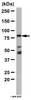
Rabbit polyclonal antibody supplied as lyophilized, undiluted serum. Recognizes the ~44-45 kDa opioid µ receptor protein.
Background
Opioid peptides are endogenous neuromodulators, which have been identified from brain, and play a major role in the nociceptive pathway by interacting with several membrane receptors, defined as µ, δ, and κ. These peptides are derived from the cleavage of precursor proteins to produce the β-endorphin, enkephalin, and dynorphin peptides. Leu-enkephalin, Met-enkephalin, and β-endorphin all bind the µ and δ receptors with high affinity, while β-endorphin also exhibits low affinity κ binding. Dynorphin A and dynorphin B both bind the κ receptor with high affinity. In addition to these endogenous peptides, a number of exogenous nonpeptide molecules known as alkaloids or opiates also interact with the receptors to mediate a number of biological events which include the modulation of pain, analgesia, behavior, locomotor activity, and neuroendocrine physiology. Although the δ opioid receptor subtype was the first subtype identified, all three receptor classes are G protein-coupled receptors which have been shown to inhibit adenylate cyclase, decrease the conductance of voltage-gated Ca2+ channels, or activate K+ channel current.
Host
Rabbit
Immunogen species
Rat
Immunogen
a synthetic peptide corresponding to amino acids 384-398 of rat opioid µ receptor
Isotype
IgG
Species
rat
Positive control
Rat caudate putamen or spinal cord (dorsal horn)
Form
Lyophilized
Formulation
Undiluted serum.
Preservative
None
Comments
The specificity was determined by immunolabeling of transfected cells, immunoblotting analysis, and immunoisolation studies. Antibody specificity was examined in the rat caudate putamen and dorsal horn of the spinal cord. Staining is completely eliminated by pre-treatment of antibody with immunogen peptide at a concentration of 10 µg/ml. This antibody has also been reported to work for immunopercipitation. Antibody should be titrated for optimal results in individual systems.
Storage
-20°C
Do Not Freeze
Ok to freeze
Special Instructions
Reconstitute the lyophilized antibody with 100 µl sterile distilled H₂O. Resulting reconstituted solution contains ≤0.1% sodium azide. Be careful to reconstitute the entire contents of the vial; during shipment and handling portions of the lyophilized pellet may have become dislodged and may not be in the bottom of the vial. Following reconstitution, aliquot and freeze (-20°C).
Toxicity
Highly Toxic
References
Zaki, P.A., et al. 1996. Annu. Rev. Pharmacol. Toxicol.36, 379. Arvidsson, U., et al. 1995. J. Neurosci.15, 3328. Kieffer, B.L. 1995. Cell. Mol. Neurobiol.15, 615-635. Childers, S.R. 1991. Life Sci.48, 1991.
Application references
Immunoblotting
Wang, X., et al. 2004. Neurosci.129, 751.
Immunoblotting, Immunofluorescence, Frozen Sections
Arvidsson, U., et al. 1995. J. Neurosci.15, 3328.


aminobenzoph[814473_-(Benzylprolyl)aminobenzoph-ALL].jpg)