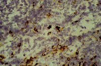
Microglial AGE-albumin is critical in promoting alcohol-induced neurodegeneration in rats and humans.
Byun, K; Bayarsaikhan, D; Bayarsaikhan, E; Son, M; Oh, S; Lee, J; Son, HI; Won, MH; Kim, SU; Song, BJ; Lee, B
PloS one
9
e104699
2014
Pokaż streszczenie
Alcohol is a neurotoxic agent, since long-term heavy ingestion of alcohol can cause various neural diseases including fetal alcohol syndrome, cerebellar degeneracy and alcoholic dementia. However, the molecular mechanisms of alcohol-induced neurotoxicity are still poorly understood despite numerous studies. Thus, we hypothesized that activated microglial cells with elevated AGE-albumin levels play an important role in promoting alcohol-induced neurodegeneration. Our results revealed that microglial activation and neuronal damage were found in the hippocampus and entorhinal cortex following alcohol treatment in a rat model. Increased AGE-albumin synthesis and secretion were also observed in activated microglial cells after alcohol exposure. The expressed levels of receptor for AGE (RAGE)-positive neurons and RAGE-dependent neuronal death were markedly elevated by AGE-albumin through the mitogen activated protein kinase pathway. Treatment with soluble RAGE or AGE inhibitors significantly diminished neuronal damage in the animal model. Furthermore, the levels of activated microglial cells, AGE-albumin and neuronal loss were significantly elevated in human brains from alcoholic indivisuals compared to normal controls. Taken together, our data suggest that increased AGE-albumin from activated microglial cells induces neuronal death, and that efficient regulation of its synthesis and secretion is a therapeutic target for preventing alcohol-induced neurodegeneration. | 25140518
 |
Immunohistochemical detection of T-cell subsets and other leukocytes in paraffin-embedded rat and mouse tissues with monoclonal antibodies.
Whiteland, J L, et al.
J. Histochem. Cytochem., 43: 313-20 (1995)
1994
Pokaż streszczenie
We describe a method for immunohistochemical localization of T-cells, CD4+ T-cells, CD8+ T-cells, B-cells, activated lymphocytes, major histocompatibility complex (MHC) class II antigens, macrophages, dendritic cells, and granulocytes in rat and mouse tissue fixed in periodate-lysine-paraformaldehyde (PLP) and embedded in paraffin. Rat and mouse spleen and eyes were fixed in PLP for 18-24 hr, rapidly dehydrated, infiltrated under vacuum with paraffin at 54 degrees C, sectioned, and stained with appropriate monoclonal antibodies (MAbs). Sections of PLP-fixed, paraffin-embedded spleen were compared with acetone-fixed frozen spleen sections with respect to morphology and staining quality. Nine of 10 MAbs to rat antigens and eight of nine MAbs to mouse antigens stained paraffin sections equally or more intensely than frozen sections. The two MAbs that showed weaker staining still gave good staining on paraffin sections. Paraffin-embedded rat and mouse eyes were easier to section serially than frozen eyes, showed superior morphology, and individually stained cells were readily identified. Therefore, a combination of PLP fixation and low-temperature paraffin embedding permits detection of the major types of immune cell in rat and mouse tissues while maintaining good morphology, particularly in diseased, damaged, or delicate tissues. | 7868861
 |


[1.00060.0100_CAYE-Broth-(100g)-ALL].jpg)


