Millipore Sigma Vibrant Logo
Attention: We have moved. Merck Millipore products are no longer available for purchase on MerckMillipore.com.Learn More

ABD16 Anti-IKAROS Antibody

ABD16
100 µg  
Purchase on Sigma-Aldrich

Oferty specjalne

Przegląd

Replacement Information

Oferty specjalne

Tabela kluczowych gatunków

Species ReactivityKey ApplicationsHostFormatAntibody Type
H, M, RWB, ICCRbAffinity PurifiedPolyclonal Antibody
Description
Catalogue NumberABD16
DescriptionAnti-IKAROS Antibody
Alternate Names
  • DNA-binding protein Ikaros
  • Ikaros family zinc finger protein 1
  • Lymphoid transcription factor LyF-1
Background InformationIKAROS, also known as IKZF1, is a transcription factor that is important for the regulation of lymphopoiesis and also the development of the immune system. It has also been linked to adult and fetal erythroid differentiation in humans. IKAROS has been shown to induce local chromatin accessibility during CD4+ T cell differentiation and therefore, activating Th2 expression.
References
Product Information
FormatAffinity Purified
Control
  • HeLa cell lysate
PresentationPurified rabbit polyclonal in buffer containing 0.1 M Tris-Glycine (pH 7.4), 150 mM NaCl with 0.05% sodium azide.
Quality LevelMQ100
Applications
ApplicationAnti-IKAROS Antibody detects level of IKAROS & has been published & validated for use in WB & IC.
Key Applications
  • Western Blotting
  • Immunocytochemistry
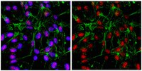
Application NotesImmunocytochemistry Analysis: 1:500 dilution from a representative lot detected IKAROS in ReNcell® human neural progenitor cells.
Biological Information
ImmunogenKLH-conjugated linear peptide corresponding to human IKAROS.
ConcentrationPlease refer to the Certificate of Analysis for the lot-specific concentration.
HostRabbit
Species Reactivity
  • Human
  • Mouse
  • Rat
Antibody TypePolyclonal Antibody
Entrez Gene Number
Gene Symbol
  • IKZF1
  • IK1
  • IKAROS
  • LYF1
  • ZNFN1A1
Purification MethodAffinity Purfied
UniProt Number
UniProt SummaryFUNCTION: Transcription regulator of hematopoietic cell differentiation. Binds gamma-satellite DNA. Binds with higher affinity to gamma satellite A. Plays a role in the development of lymphocytes, B- and T-cells. Binds and activates the enhancer (delta-A element) of the CD3-delta gene. Repressor of the TDT (terminal deoxynucleotidyltransferase) gene during thymocyte differentiation. Regulates transcription through association with both HDAC-dependent and HDAC-independent complexes. Targets the 2 chromatin-remodeling complexes, NuRD and BAF (SWI/SNF), in a single complex (PYR complex), to the beta-globin locus in adult erythrocytes. Increases normal apoptosis in adult erythroid cells. Confers early temporal competence to retinal progenitor cells (RPCs) (By similarity).

SUBUNIT STRUCTURE: Heterodimer with other IKAROS family members. Interacts with IKZF4 AND IKZF5 By similarity. Component of the chromatin-remodeling NuRD repressor complex which includes at least HDAC1, HDAC2, RBBP4, RBBP7, IKZF1, MTA2, MBD2, MBD3, MTA1L1, CHD3 and CHD4. Interacts directly with the CHD4 component of the NuRD complex. Component of the BAF (SWI/SNF) gene activator complex which includes ACTB, ARID1A, ARID1B, IKZF1, ARID1A, ARID1B, SMARCA2, SMARCA4 and at least one BAF subunit. Interacts directly with the SMARCA4 component of the BAF complex (By similarity). Interacts with SUMO1; the interaction sumoylates IKAROS, promoted by PIAS2 and PIAS3. Interacts with PIAS2 (isoform alpha); the interaction promotes sumoylation and reduces transcription repression. Interacts, to a lesser extent, with PIAS3. Interacts with PPP1CC; the interaction targets PPP1CC to pericentromeric heterochromatin, dephosphorylates IKAROS, stabilizes it and prevents it from degradation (By similarity).

SUBCELLULAR LOCATION: Nucleus. Note: In resting lymphocytes, distributed diffusely throughout the nucleus. Localizes to pericentromeric heterochromatin in proliferating cells. This localization requires DNA binding which is regulated by phosphorylation / dephosphorylation events (By similarity).

Isoform Ik2: Nucleus. Note: In resting lymphocytes, distributed diffusely throughout the nucleus. Localizes to pericentromeric heterochromatin in proliferating cells. This localization requires DNA binding which is regulated by phosphorylation / dephosphorylation events (By similarity).

Isoform Ik6: Cytoplasm (By similarity).

TISSUE SPECIFICTY: Abundantly expressed in thymus, spleen and peripheral blood Leukocytes and lymph nodes. Lower expression in bone marrow and small intestine.

DOMAIN: The N-terminal zinc-fingers 2 and 3 are required for DNA binding as well as for targeting IKFZ1 to pericentromeric heterochromatin (By similarity).

The C-terminal zinc-finger domain is required for dimerization (By similarity).

PTM: Phosphorylation controls cell-cycle progression from late G1 stage to S stage. Hyperphosphorylated during G2/M phase. Dephosphorylated state during late G1 phase. Phosphorylation on Thr-140 is required for DNA and pericentromeric location during mitosis. CK2 is the main kinase, in vitro. GSK3 and CDK may also contribute to phosphorylation of the C-terminal serine and threonine residues. Phosphorylation on these C-terminal residues reduces the DNA-binding ability. Phosphorylation/dephosphorylation events on Ser-13 and Ser-295 regulate TDT expression during thymocyte differentiation. Dephosphorylation by protein phosphatase 1 regulates stability and pericentromeric heterochromatin location. Phosphorylated in both lymphoid and non-lymphoid tissues (By similarity).

Sumoylated. Simulataneous sumoylation on the 2 sites results in a loss of both HDAC-dependent and HDAC-independent repression. Has no effect on pericentromeric heterochromatin location. Desumoylated by SENP1 (By similarity).

Polyubiquitinated (By similarity).

INVOLVEMENT IN DISEASE: Note=Defects in IKZF1 are frequent occurrences (28.6%) in acute lymphoblasic leukemia (ALL). Such alterations or deletions lead to poor prognosis for ALL.

SEQUENCE SIMILARITIES: Belongs to the Ikaros C2H2-type zinc-finger protein family.

Contains 6 C2H2-type zinc fingers.
Molecular Weight~65 kDa observed. (Martin-Ibanez, R., et al. (2010). The Journal of Comparative Neurology. 518:329–351.) An uncharacterized band appears at ~57 kDa in some lysates.
Physicochemical Information
Dimensions
Materials Information
Toxicological Information
Safety Information according to GHS
Safety Information
Product Usage Statements
Quality AssuranceEvaluated by Western Blot in HeLa cell lysate.

Western Blot Analysis: 0.5 µg/mL of this antibody detected IKAROS on 10 µg of HeLa cell lysate.
Usage Statement
  • Unless otherwise stated in our catalog or other company documentation accompanying the product(s), our products are intended for research use only and are not to be used for any other purpose, which includes but is not limited to, unauthorized commercial uses, in vitro diagnostic uses, ex vivo or in vivo therapeutic uses or any type of consumption or application to humans or animals.
Storage and Shipping Information
Storage ConditionsStable for 1 year at 2-8°C from date of receipt.
Packaging Information
Material Size100 µg
Transport Information
Supplemental Information
Specifications
Global Trade Item Number
Numer katalogowy GTIN
ABD16 04053252527524