


NS225 Sigma-AldrichNeurite Outgrowth Assay Kit (1 µm)

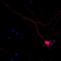
The NS225 Neurite Outgrowth Assay Kit (1 μm) is based on the use of Millicell cell culture inserts (chambers) containing a permeable membrane with 1 μm pores at the base.
More>> The NS225 Neurite Outgrowth Assay Kit (1 μm) is based on the use of Millicell cell culture inserts (chambers) containing a permeable membrane with 1 μm pores at the base. Less<<Neurite Outgrowth Assay Kit (1 µm) MSDS (material safety data sheet) or SDS, CoA and CoQ, dossiers, brochures and other available documents.
Recommended Products
Overview
| Replacement Information |
|---|
Key Spec Table
| Detection Methods |
|---|
| Chromogenic |
| References |
|---|
| Product Information | |
|---|---|
| Components |
|
| Detection method | Chromogenic |
| HS Code | 3822 19 90 |
| Quality Level | MQ100 |
| Applications | |
|---|---|
| Application | The NS225 Neurite Outgrowth Assay Kit (1 μm) is based on the use of Millicell cell culture inserts (chambers) containing a permeable membrane with 1 μm pores at the base. |
| Biological Information | |
|---|---|
| Species Reactivity |
|
| Physicochemical Information |
|---|
| Dimensions |
|---|
| Materials Information |
|---|
| Toxicological Information |
|---|
| Safety Information according to GHS |
|---|
| Safety Information |
|---|
| Packaging Information | |
|---|---|
| Material Size | 12 assays |
| Transport Information |
|---|
| Supplemental Information |
|---|
| Specifications |
|---|
| Global Trade Item Number | |
|---|---|
| Catalogue Number | GTIN |
| NS225 | 04053252660085 |
Documentation
Neurite Outgrowth Assay Kit (1 µm) SDS
| Title |
|---|
References
| Reference overview | Pub Med ID |
|---|---|
| Neural progenitor cells derived from adult bone marrow mesenchymal stem cells promote neuronal regeneration. Yue Tang,Yong-Chun Cui,Xiao-Juan Wang,Ai-Li Wu,Guang-Fu Hu,Fu-Liang Luo,Jia-Kang Sun,Jing Sun,Li-Ke Wu Life sciences 91 2012 Show Abstract | 23000028
|
| Small GTPase Tc10 and its homologue RhoT induce N-WASP-mediated long process formation and neurite outgrowth. Abe, Tomoyuki, et al. J. Cell. Sci., 116: 155-68 (2003) 2003 Show Abstract | 12456725
|
| Spinal cord repair: strategies to promote axon regeneration. McKerracher, L Neurobiol. Dis., 8: 11-8 (2001) 2001 Show Abstract | 11162236
|
| Repulsive factors and axon regeneration in the CNS Fournier, A E and Strittmatter, S M Curr Opin Neurobiol, 11:89-94 (2001) 2001 | 11179877
|
| Glial inhibition of nerve regeneration in the mature mammalian CNS. Qiu, J, et al. Glia, 29: 166-74 (2000) 2000 Show Abstract | 10625335
|
| Axon regeneration: Vaccinating against spinal cord injury Filbin, M T Curr Biol, 10:R100-R103 (2000) 2000 | 10679313
|
| Extracellular matrix allows PC12 neurite elongation in the absence of microtubules Lamoureux, P, et al. J Cell Biol, 110:71-79 (1990) 1990 | 2153148
|
| Nerve growth factor-induced neurite outgrowth in PC12 cells involves the coordinate induction of microtubule assembly and assembly-promoting factors Drubin, D G, et al J Cell Biol, 101:1799-1807 (1985) 1985 | 2997236
|
Related Products & Applications
Product Families
Nervous System MarkersThe nervous system coordinates the voluntary and involuntary actions of the individual and transmits signals between different parts of the body.Learn More >> |

Migration and Invasion AssaysEMD Millipore offers a wide array of migration, invasion, chemotactic and haptotactic Boyden Chamber assays for cell migration studies.Learn More >> |
Categories
| Life Science Research > Cell Analysis > Cell-based Assays > Neurite Outgrowth |