Millipore Sigma Vibrant Logo
Attention: We have moved. Merck Millipore products are no longer available for purchase on MerckMillipore.com.Learn More

17-10245 ChIPAb+™ Histone H3.3 - ChIP Validated Antibody and Primer Set

View Products on Sigmaaldrich.com
17-10245
25 assays  25 assays per set. Recommended use: ~2 μg of antibody per chromatin immunoprecipitation (dependent upon biological context).
Purchase on Sigma-Aldrich

Special Offers

Overview

Replacement Information

Key Spec Table

Species ReactivityKey Applications
H, MDB, WB, ChIP
Description
Catalogue Number17-10245
Trade Name
  • ChIPAb+
  • Upstate
DescriptionChIPAb+™ Histone H3.3 - ChIP Validated Antibody and Primer Set
OverviewAll ChIPAb+ antibodies are individually validated for chromatin precipitation, every lot, every time. Each ChIPAb+ antibody set includes control primers (tested every lot by qPCR) to biologically validate your IP results in a locus-specific context. The qPCR protocol and primer sequences are provided, allowing researchers to validate ChIP protocols when using our antibody in their chromatin context. Each set also includes a negative control antibody to ensure specificity of the ChIP reaction.
The ChIPAb+ Histone H3.3 set includes the Histone H3.3 antibody, a Normal rabbit IgG, and control primers which amplify a 87 bp region of ChIP Primers, GAPDH Coding Region D2 Human. The Histone H3.3 and negative controls are supplied in a scalable "per ChIP" reaction size and can be used to functionally validate the precipitation of Histone H3.3 -associated chromatin.
Alternate Names
  • Histone H3.3
Background InformationHistone H3 is one of the five main histone proteins involved in the structure of chromatin in eukaryotic cells. Featuring a main globular domain and a long N-terminal tail, H3 is involved with the structure of the nucleosomes of the 'beads on a string' structure. The N-terminal tail of histone H3 protrudes from the globular nucleosome core and can undergo several different types of epigenetic modifications that influence cellular processes. These modifications include the covalent attachment of methyl or acetyl groups to lysine and arginine amino acids and the phosphorylation of serine or threonine. Histone variant H3.3 is typically enriched in active chromatin.
References
Product Information
FormatAffinity Purified
Control
  • Includes normal rabbit IgG and primers specific for human GAPDH coding region.
PresentationAnti-Histone H3.3 (rabbit polyclonal). One vial containing 50 µg of purified rabbit polyclonal in buffer containing 0.1M Tris-Glycine (pH 7.4), 150 mM NaCl with 0.05% sodium azide before the addition of glycerol to 30%. Store at -20° C.
Concentration: 0.7 mg/mL
Normal Rabbit IgG. One vial containing 125 µg of Rabbit IgG in 125 µL of storage buffer containing 0.05% sodium azide. Store at -20°C.
Control Primers, GAPDH Coding Region D2 Human. One vial containing 75 μL of 5 μM of each primer specific for human GAPDH coding region. Store at -20°C.
FOR: GCC ATG TAG ACC CCT TGA AGA G
REV: ACT GGT TGA GCA CAG GGT ACT TTA T
Quality LevelMQ100
Applications
ApplicationThis ChIPAb+ Histone H3.3 -ChIP Validated Antibody & Primer Set conveniently includes the antibody & the specific control PCR primers.
Key Applications
  • Dot Blot
  • Western Blotting
  • Chromatin Immunoprecipitation (ChIP)
Application NotesChromatin Immunoprecipitation:
Representative lot data.
Sonicated chromatin prepared from HeLa cells (1 X 10E6 cell equivalents per IP) were subjected to chromatin immunoprecipitation using 2 µg of either Normal rabbit IgG or 2 µg Anti-Histone H3.3 and the Magna ChIP™ A Kit (Cat. # 17-610). Successful immunoprecipitation of Histone H3.3 associated DNA fragments was verified by qPCR using ChIP Primers, GAPDH Coding Region D2 as a positive locus, and beta-actin promoter primers as a negative locus. (Figure 2). Data is presented as percent input of each IP sample relative to input chromatin for each amplicon and ChIP sample as indicated.
Please refer to the EZ-Magna ChIP™ A (Cat. # 17-408) or EZ-ChIP™ (Cat. # 17-371) protocol for experimental details.
Western Blot Analysis:
Representative lot data.
HeLa acid extract was probed with Anti-Histone H3.3 (1 µg/mL). Proteins were visualized using a Donkey Anti-Rabbit IgG secondary antibody conjugated to HRP and a chemiluminescence detection system.
Arrow indicates Histone H3.3 (~17 kDa). (Figure 3).
Dot Blot Analysis;
Representative lot data.
Recombinant H3.2 and H3.3 spotted on PVDF (1, 10, 100, 1,000 ng histone per slot). Detection with 0.5 µg/mL a-H3.3 antibody in TBS-T, 5% milk. (Figure 4).
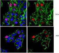
Image courtesy of Simon Elsässer, Laboratory of David Allis, Rockefeller University, New York.
Biological Information
ImmunogenKLH-conjugated linear peptide corresponding to human Histone H3.3.
Epitopea.a. 85-105
ConcentrationPlease refer to the Certificate of Analysis for the lot-specific concentration.
HostRabbit
SpecificityThis antibody recognizes human Histone H3.
Species Reactivity
  • Human
  • Mouse
Species Reactivity NoteDemonstrated to react with Human and Mouse. Broad species cross-reactivity expected based on 100% sequence homology.
Antibody TypePolyclonal Antibody
Entrez Gene Number
Entrez Gene SummaryHistones are basic nuclear proteins that are responsible for the nucleosome structure of the chromosomal fiber in eukaryotes. Two molecules of each of the four core histones (H2A, H2B, H3, and H4) form an octamer, around which approximately 146 bp of DNA is wrapped in repeating units, called nucleosomes. The linker histone, H1, interacts with linker DNA between cleosomes and functions in the compaction of chromatin into higher order structures. This gene contains introns and its mRNA is polyadenylated, unlike most histone genes. The protein encoded is a replication-independent member of the histone H3 family. [provided by RefSeq].
Gene Symbol
  • H3F3A
  • H3.3A
  • H3F3
  • PP781
  • H3F3B
  • H3.3B
Purification MethodAffinity Purfied
UniProt Number
UniProt SummaryFUNCTION: Variant histone H3 which replaces conventional H3 in a wide range of nucleosomes in active genes. Constitutes the predominant form of histone H3 in non-dividing cells and is incorporated into chromatin independently of DNA synthesis. Deposited at sites of nucleosomal displacement throughout transcribed genes, suggesting that it represents an epigenetic imprint of transcriptionally active chromatin. Nucleosomes wrap and compact DNA into chromatin, limiting DNA accessibility to the cellular machineries which require DNA as a template. Histones thereby play a central role in transcription regulation, DNA repair, DNA replication and chromosomal stability. DNA accessibility is regulated via a complex set of post-translational modifications of histones, also called histone code, and nucleosome remodeling.

SUBUNIT STRUCTURE: The nucleosome is a histone octamer containing two molecules each of H2A, H2B, H3 and H4 assembled in one H3-H4 heterotetramer and two H2A-H2B heterodimers. The octamer wraps approximately 147 bp of DNA. Interacts with HIRA, a chaperone required for its incorporation into nucleosomes.

SUBCELLULAR LOCATION: Nucleus.

DEVELOPMENTAL STAGE: Expressed throughout the cell cycle independently of DNA synthesis.

PTM: Acetylation is generally linked to gene activation. Acetylation on Lys-10 (H3K9ac) impairs methylation at Arg-9 (H3R8sme2). Acetylation on Lys-19 (H3K18ac) and Lys-24 (H3K24ac) favors methylation at Arg-18 (H3R17me).

Citrullination at Arg-9 (H3R8ci) and/or Arg-18 (H3R17ci) by PADI4 impairs methylation and represses transcription.

Asymmetric dimethylation at Arg-18 (H3R17me2a) by CARM1 is linked to gene activation. Symmetric dimethylation at Arg-9 (H3R8sme2) by PRMT5 is linked to gene repression. Asymmetric dimethylation at Arg-3 (H3R2me2a) by PRMT6 is linked to gene repression and is mutually exclusive with H3 Lys-5 methylation (H3K4me2 and H3K4me3). H3R2me2a is present at the 3' of genes regardless of their transcription state and is enriched on inactive promoters, while it is absent on active promoters.

Specifically enriched in modifications associated with active chromatin such as methylation at Lys-5 (H3K4me), Lys-37 and Lys-80. Methylation at Lys-5 (H3K4me) facilitates subsequent acetylation of H3 and H4. Methylation at Lys-80 (H3K79me) is associated with DNA double-strand break (DSB) responses and is a specific target for TP53BP1. Methylation at Lys-10 (H3K9me) and Lys-28 (H3K27me), which are linked to gene repression, are underrepresented. Methylation at Lys-10 (H3K9me) is a specific target for HP1 proteins (CBX1, CBX3 and CBX5) and prevents subsequent phosphorylation at Ser-11 (H3S10ph) and acetylation of H3 and H4. Methylation at Lys-5 (H3K4me) and Lys-80 (H3K79me) require preliminary monoubiquitination of H2B at 'Lys-120'. Methylation at Lys-10 (H3K9me) and Lys-28 (H3K27me) are enriched in inactive X chromosome chromatin.

Phosphorylated at Thr-4 (H3T3ph) by GSG2/haspin during prophase and dephosphorylated during anaphase. At centromeres, specifically phosphorylated at Thr-12 (H3T11ph) from prophase to early anaphase, probably DAPK3. Phosphorylation at Ser-11 (H3S10ph) by AURKB is crucial for chromosome condensation and cell-cycle progression during mitosis and meiosis. In addition phosphorylation at Ser-11 (H3S10ph) by RPS6KA4 and RPS6KA5 is important during interphase because it enables the transcription of genes following external stimulation, like mitogens, stress, growth factors or UV irradiation and result in the activation of genes, such as c-fos and c-jun. Phosphorylation at Ser-11 (H3S10ph), which is linked to gene activation, prevents methylation at Lys-10 (H3K9me) but facilitates acetylation of H3 and H4. Phosphorylation at Ser-11 (H3S10ph) by AURKB mediates the dissociation of HP1 proteins (CBX1, CBX3 and CBX5) from heterochromatin. Phosphorylation at Ser-11 (H3S10ph) is also an essential regulatory mechanism for neoplastic cell transformation. Phosphorylated at Ser-29 by MLTK isoform 1, RPS6KA5 or AURKB during mitosis or upon ultraviolet B irradiation. Phosphorylation on Ser-32 is specific to regions bordering centromeres in metaphase chromosomes.

Ubiquitinated (By similarity).

SEQUENCE SIMILARITIES: Belongs to the histone H3 family.

SEQUENCE CAUTION: The sequence CAH73371.1 differs from that shown. Reason: Erroneous gene model prediction.
Molecular Weight~17 kDa observed
Physicochemical Information
Dimensions
Materials Information
Toxicological Information
Safety Information according to GHS
Safety Information
Product Usage Statements
Quality AssuranceChromatin Immunoprecipitation:
Representative lot data.
Sonicated chromatin prepared from HeLa cells (1 X 10E6 cell equivalents per IP) were subjected to chromatin immunoprecipitation using 2 µg of either Normal Rabbit IgG or 2 µg of Anti-Histone H3.3 and the Magna ChIP™ A Kit (Cat. # 17-610). Successful immunoprecipitation of Histone H3.3 associated DNA fragments was verified by qPCR using ChIP Primers, GAPDH Coding Region D2 (Figure 1).
Please refer to the EZ-Magna ChIP™ A (Cat. # 17-408) or EZ-ChIP™ (Cat. # 17-371) protocol for experimental details.
Usage Statement
  • Unless otherwise stated in our catalog or other company documentation accompanying the product(s), our products are intended for research use only and are not to be used for any other purpose, which includes but is not limited to, unauthorized commercial uses, in vitro diagnostic uses, ex vivo or in vivo therapeutic uses or any type of consumption or application to humans or animals.
Storage and Shipping Information
Storage ConditionsStable for 1 year at -20°C from date of receipt. Handling Recommendations: Upon first thaw, and prior to removing the cap, centrifuge the vial and gently mix the solution. Aliquot into microcentrifuge tubes and store at -20°C. Avoid repeated freeze/thaw cycles, which may damage IgG and affect product performance.
Note: Variability in freezer temperatures below -20°C may cause glycerol containing solutions to become frozen during storage.
Packaging Information
Material Size25 assays
Material Package25 assays per set. Recommended use: ~2 μg of antibody per chromatin immunoprecipitation (dependent upon biological context).
Transport Information
Supplemental Information
Specifications
Global Trade Item Number
Catalogue Number GTIN
17-10245 04053252293474

Documentation

ChIPAb+™ Histone H3.3 - ChIP Validated Antibody and Primer Set SDS

Title

Safety Data Sheet (SDS) 

ChIPAb+™ Histone H3.3 - ChIP Validated Antibody and Primer Set Certificates of Analysis

TitleLot Number
ChIPAb+ Histone H3.3 - 1986165 1986165
ChIPAb+ Histone H3.3 - 2022054 2022054
ChIPAb+ Histone H3.3 - 2060988 2060988
ChIPAb+ Histone H3.3 - 2196084 2196084
ChIPAb+ Histone H3.3 - NRG1915763 NRG1915763
ChIPAb+™ Histone H3.3 2471014
ChIPAb+™ Histone H3.3 - 3204328 3204328
ChIPAb+™ Histone H3.3 - 3452518 3452518
ChIPAb+™ Histone H3.3 - 3698771 3698771
ChIPAb+™ Histone H3.3 - 3756642 3756642

References

Reference overviewPub Med ID
Analysis of neonatal brain lacking ATRX or MeCP2 reveals changes in nucleosome density, CTCF binding and chromatin looping.
Kernohan, KD; Vernimmen, D; Gloor, GB; Bérubé, NG
Nucleic acids research  42  8356-68  2014

Show Abstract
24990380 24990380

Brochure

Title
New Products: Volume 3, 2012

Related Products & Applications

Product Families

Related Products By: Brand Facete

Categories

Life Science Research > Antibodies and Assays > Immunoassays > Immunoprecipitation (IP) > Chromatin Immunoprecipitation (ChIP) > ChIP Validated Antibodies
Life Science Research > Antibodies and Assays > Primary Antibodies
Life Science Research > Protein Detection and Quantification > Immunoassays > Immunoprecipitation (IP) > Chromatin Immunoprecipitation (ChIP) > ChIP Validated Antibodies