Our broad portfolio consists of multiplex panels that allow you to choose, within the panel, analytes that best meet your needs. On a separate tab you can choose the premixed cytokine format or a single plex kit.
Cell Signaling Kits & MAPmates™
Choose fixed kits that allow you to explore entire pathways or processes. Or design your own kits by choosing single plex MAPmates™, following the provided guidelines.
The following MAPmates™ should not be plexed together:
-MAPmates™ that require a different assay buffer
-Phospho-specific and total MAPmate™ pairs, e.g. total GSK3β and GSK3β (Ser 9)
-PanTyr and site-specific MAPmates™, e.g. Phospho-EGF Receptor and phospho-STAT1 (Tyr701)
-More than 1 phospho-MAPmate™ for a single target (Akt, STAT3)
-GAPDH and β-Tubulin cannot be plexed with kits or MAPmates™ containing panTyr
.
Catalogue Number
Ordering Description
Qty/Pack
List
This item has been added to favorites.
Select A Species, Panel Type, Kit or Sample Type
To begin designing your MILLIPLEX® MAP kit select a species, a panel type or kit of interest.
Custom Premix Selecting "Custom Premix" option means that all of the beads you have chosen will be premixed in manufacturing before the kit is sent to you.
Catalogue Number
Ordering Description
Qty/Pack
List
This item has been added to favorites.
Species
Panel Type
Selected Kit
Qty
Catalogue Number
Ordering Description
Qty/Pack
List Price
96-Well Plate
Qty
Catalogue Number
Ordering Description
Qty/Pack
List Price
Add Additional Reagents (Buffer and Detection Kit is required for use with MAPmates)
Qty
Catalogue Number
Ordering Description
Qty/Pack
List Price
48-602MAG
Buffer Detection Kit for Magnetic Beads
1 Kit
Space Saver Option Customers purchasing multiple kits may choose to save storage space by eliminating the kit packaging and receiving their multiplex assay components in plastic bags for more compact storage.
This item has been added to favorites.
The Product Has Been Added To Your Cart
You can now customize another kit, choose a premixed kit, check out or close the ordering tool.
Attention: We have moved. Merck Millipore products are no longer available for purchase on MerckMillipore.com.Learn More
345860
Sigma-AldrichAnti-Glial Fibrillary Acidic Protein Rat mAb (2.2B10)
This Anti-Glial Fibrillary Acidic Protein Rat mAb (2.2B10) is validated for use in ELISA, Immunoblotting, Frozen Sections, IP, Paraffin Sections for the detection of Glial Fibrillary Acidic Protein.
More>>This Anti-Glial Fibrillary Acidic Protein Rat mAb (2.2B10) is validated for use in ELISA, Immunoblotting, Frozen Sections, IP, Paraffin Sections for the detection of Glial Fibrillary Acidic Protein. Less<<
MSDS (material safety data sheet) or SDS, CoA and CoQ, dossiers, brochures and other available documents.
Recognizes GFAP in astrocytes induced by a variety of CNS injuries in human and rat brain tissues.
Catalogue Number
345860
Brand Family
Calbiochem®
Synonyms
Anti-GFAP
Application Data
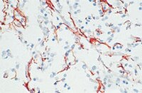
Detection of human glial fibrillary acidic protein by immunohistochemistry. Sample: Astrocytoma tissue. Primary antibody: Anti-Glial Fibrillary Acidic Protein Rat mAb (2.2B10) (Cat. No. 345860) (10 µg/ml). Detection: fluorescence.
References
References
Tohyama, T., et al. 1993. Am. J. Pathol.142, 871. Lee, V.M., et al. 1984. J. Neurochem. 42, 25.
Product is not to be used for animal treatment, in vivo research, or in any other contact procedure with livestock. Stains reactive rodent and human brain astrocytes induced by a variety of central nervous system injuries. This antibody is suitable for immunohistochemical staining of Bouin's-fixed, frozen, or paraffin-embedded tissue sections. Variables associated with assay conditions will dictate the proper working dilution.
Biological Information
Immunogen
bovine GFAP
Immunogen
Bovine Brain
Clone
2.2B10
Host
Rat
Isotype
IgG2a
Species Reactivity
Bovine
Human
Mouse
Rat
Antibody Type
Monoclonal Antibody
Concentration Label
Please refer to vial label for lot-specific concentration
Storage and Shipping Information
Ship Code
Blue Ice Only
Toxicity
Standard Handling
Storage
-20°C
Avoid freeze/thaw
Avoid freeze/thaw
Do not freeze
Ok to freeze
Special Instructions
Following initial thaw, aliquot and freeze (-20°C).
Global Trade Item Number
Catalogue Number
GTIN
345860-100UG
04055977214413
Documentation
Anti-Glial Fibrillary Acidic Protein Rat mAb (2.2B10) SDS
Anti-Glial Fibrillary Acidic Protein Rat mAb (2.2B10) Certificates of Analysis
Title
Lot Number
345860
References
Reference overview
Tohyama, T., et al. 1993. Am. J. Pathol.142, 871. Lee, V.M., et al. 1984. J. Neurochem. 42, 25.
Data Sheet
Note that this data sheet is not lot-specific and is representative of the current specifications for this product. Please consult the vial label and the certificate of analysis for information on specific lots. Also note that shipping conditions may differ from storage conditions.
Detection of human glial fibrillary acidic protein by immunohistochemistry. Sample: Astrocytoma tissue. Primary antibody: Anti-Glial Fibrillary Acidic Protein Rat mAb (2.2B10) (Cat. No. 345860) (10 µg/ml). Detection: fluorescence.
Description
Purified rat monoclonal antibody. Recognizes the ~55 kDa glial fibrillary acidic protein (GFAP).
Host
Rat
Immunogen species
Bovine Brain
Immunogen
bovine GFAP
Clone
2.2B10
Isotype
IgG2a
Species
bovine, human, rodent
Positive control
Human or rat brain tissues
Form
Liquid
Formulation
In PBS.
Concentration Label
Please refer to vial label for lot-specific concentration
Preservative
≤0.1% sodium azide
Comments
Product is not to be used for animal treatment, in vivo research, or in any other contact procedure with livestock. Stains reactive rodent and human brain astrocytes induced by a variety of central nervous system injuries. This antibody is suitable for immunohistochemical staining of Bouin's-fixed, frozen, or paraffin-embedded tissue sections. Variables associated with assay conditions will dictate the proper working dilution.
Storage
Avoid freeze/thaw -20°C
Do Not Freeze
Ok to freeze
Special Instructions
Following initial thaw, aliquot and freeze (-20°C).
Toxicity
Standard Handling
References
Tohyama, T., et al. 1993. Am. J. Pathol.142, 871. Lee, V.M., et al. 1984. J. Neurochem. 42, 25.