Millipore Sigma Vibrant Logo
Attention: We have moved. Merck Millipore products are no longer available for purchase on MerckMillipore.com.Learn More

CBL496-I-25UL Anti-CD34 Antibody, clone QBEnd/10

View Products on Sigmaaldrich.com
CBL496-I-25UL
25 µL  
Purchase on Sigma-Aldrich

Overview

Replacement Information

Key Spec Table

Species ReactivityKey ApplicationsHostFormatAntibody Type
Mk, HEM, FC, IF, FACS, IH(P), WBMPurifiedMonoclonal Antibody
Description
Catalogue NumberCBL496-I-25UL
DescriptionAnti-CD34 Antibody, clone QBEnd/10
Alternate Names
  • Hematopoietic progenitor cell antigen CD34
Background InformationHematopoietic progenitor cell antigen CD34 (UniProt: P28906; also known as CD34) is encoded by the CD34 gene (Gene ID: 947) in human. CD34 is a highly glycosylated single-pass type I membrane protein that is expressed on hematopoietic progenitor cells and small vessel endothelium of a variety of tissues. Under normal conditions, CD34+ expressing cells account for about 1 2% of the total bone marrow cells. It serves as an adhesion molecule that plays a role in early hematopoiesis by mediating the attachment of stem cells to the bone marrow extracellular matrix or directly to stromal cells. It is also reported to act as a scaffold for the attachment of lineage specific glycans, allowing stem cells to bind to lectins expressed by stromal cells or other marrow components. CD34 is synthesized with a signal peptide (aa 1-31) that is cleaved off in the mature form. The mature form has an extracellular domain (aa 32-290), a transmembrane domain (aa 291-311), and a cytoplasmic domain (aa 312-385). Two isoforms of CD34 have been described that are produced by alternative splicing.
References
Product Information
FormatPurified
PresentationPurified mouse monoclonal antibody IgG1 in buffer containing 0.1 M Tris-Glycine (pH 7.4), 150 mM NaCl with 0.05% sodium azide.
Applications
Application
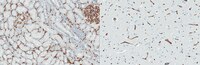
Anti-CD34, clone QBEnd/10, Cat. No. CBL496-I, is a mouse monoclonal antibody that detects CD34 and has been tested for use in Electron Microscopy, Flow Cytometry, Immunofluorescence and Fluorescence Activated Cell Sorting (FACS), Immunohistochemistry (Paraffin), and Western Blotting.
Key Applications
  • Electron Microscopy
  • Flow Cytometry
  • Immunofluorescence
  • Fluorescence Activated Cell Sorting (FACS)
  • Immunohistochemistry (Paraffin)
  • Western Blotting
Application NotesImmunohistochemistry (Paraffin) Analysis: A 1:250 dilution from a representative lot detected CD34 in human brain tissue sections.

Fluorescence Activated Cell Sorting (FACS) Analysis: A representative lot was used to sort CD34+ cells from bone marrow. (de Bock, C.E., et. al. (2012). Leukemia. 26(5):918-26).

Immunofluorescence Analysis: A representative lot detected CD34 in Immunofluorescence applications (Miki, T., et. al. (2010). Mol Cancer Res. 8(5):665-76).

Electron Microscopy Analysis: A representative lot detected CD34 in Electron Microscopy applications (Fina, L., et. al. (1990). Blood. 75(12):2417-26).

Immunohistochemistry Analysis: A representative lot detected CD34 in Immunohistochemistry applications (Engler, J.R., et. al. (2012). PLoS One. 7(8):e43339; Fina, L., et. al. (1990). Blood. 75(12):2417-26).

Flow Cytometry Analysis: A representative lot detected CD34 in Flow Cytometry applications (de Bock, C.E., et. al. (2012). Leukemia. 26(5):918-26; Fina, L., et. al. (1990). Blood. 75(12):2417-26).

Western Blotting Analysis: A representative lot detected CD34 in Western Blotting applications (Fina, L., et. al. (1990). Blood. 75(12):2417-26).
Biological Information
ImmunogenHuman placental endothelial membrane vesicles.
CloneQBEnd/10
ConcentrationPlease refer to lot specific datasheet.
HostMouse
SpecificityClone QBEnd/10 specifically detects CD34 in human and non-human primates.
IsotypeIgG1λ
Species Reactivity
  • Monkey
  • Human
Species Reactivity NoteHuman, Monkey.
Antibody TypeMonoclonal Antibody
Entrez Gene Number
Gene Symbol
  • CD34
Purification MethodProtein G purified
UniProt Number
Molecular Weight40.72 kDa Calculated. This antibody recognizes a heavily glycosylated transmembrane protein: gp 105-120 kDa.
Physicochemical Information
Dimensions
Materials Information
Toxicological Information
Safety Information according to GHS
Safety Information
Product Usage Statements
Quality AssuranceEvaluated by Immunohistochemistry (Paraffin) in human kidney tissue sections.

Immunohistochemistry (Paraffin) Analysis: A 1:250 dilution of this antibody detected CD34 in human kidney tissue sections.
Usage Statement
  • Unless otherwise stated in our catalog or other company documentation accompanying the product(s), our products are intended for research use only and are not to be used for any other purpose, which includes but is not limited to, unauthorized commercial uses, in vitro diagnostic uses, ex vivo or in vivo therapeutic uses or any type of consumption or application to humans or animals.
Storage and Shipping Information
Storage ConditionsStable for 1 year at -20°C from date of receipt. Handling Recommendations: Upon receipt and prior to removing the cap, centrifuge the vial and gently mix the solution. Aliquot into microcentrifuge tubes and store at -20°C. Avoid repeated freeze/thaw cycles, which may damage IgG and affect product performance.
Packaging Information
Material Size25 µL
Transport Information
Supplemental Information
Specifications
Global Trade Item Number
Catalogue Number GTIN
CBL496-I-25UL 04054839577109