C100906Stable gradient for quantitative, real-time, slide-based assays
Traditional multiwell (Boyden chamber) migration assays can have limitations, such as: (1) gradients are not linear, well-established, or controlled, (2) quantification of migratory cells is based on an endpoint measurement rather than a dynamic determination, and (3) imaging of the cells while they are migrating is currently not possible.
The Millicell µ-Migration Assay Kit overcomes these limitations.
Its microfluidic, low-volume technology promotes a stable, diffusion-generated concentration gradient that is consistently linear and lasts for more than 48 hours. Designed for video microscopy assays, the µ-Migration Slide is made from a plastic with high optical qualities similar to those of glass. At specific time intervals, images of the observation area can be acquired, allowing real-time monitoring and quantitative measurements of cell migration.
Benefits
- Live cell tracking of single cell or cell population migration
- Establishes a stable and linear concentration gradient that lasts ≥ 48 hours
- Helps distinguish chemotaxis from random movement
- Allows for multiparametric analysis, including cell directionality and velocity
- Enhanced optical imaging of slow- and fast-migrating cells
- Analyze 3 chemoattractants in parallel for increased throughput and flexibility
- No need to spend time or money on optimization
- Ready to use, no assembly needed
How the Kit Works
For detailed videos and instructions on the use of the µ-migration kit, please visit: Millicell® µ-Migration Assay Kit
For instructions on how to access and use the free Image-J plug-in software to analyze the data and images collected using the µ-Migration Assay Kit, please visit: Millicell® µ-Migration Assay Kit.
Supporting Data
Move your research and your cells forward
A stable, linear concentration gradient
The concentration profile remains linear over the 1 mm observation window after 1 hour and is linear and stable for over 48 hours.
Image examples of flourescence measurements

Relative intensity across a 1 mm slit (raw and smoothed data)
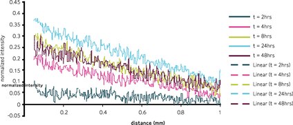
**The calculated maximum concentration is only 70% of the applied concentration. The final maximum concentration is only 33% of the applied concentration. Compare this with a Boyden chamber where very steep gradients may form along a single axis perpendicular to the surface of the membrane.
Compatibility with multiple cell lines
HT-1080, HUVEC, MDA-MB-231, and NIH-3T3 cell lines have been shown to work effectively with the Millicell µ-migration kit.


)
)
)