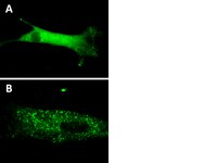
17-10224 Sigma-AldrichLentiBrite™ GFP-p62 Lentiviral Biosensor
Recommended Products
Overview
| Replacement Information |
|---|
Key Spec Table
| Key Applications | Detection Methods |
|---|---|
| TFX, IF, ICC | Fluorescent |
| Description | |
|---|---|
| Catalogue Number | 17-10224 |
| Trade Name |
|
| Description | LentiBrite™ GFP-p62 Lentiviral Biosensor |
| Overview | Read our application note in Nature Methods! http://www.nature.com/app_notes/nmeth/2012/121007/pdf/an8620.pdf (Click Here!) Learn more about the advantages of our LentiBrite Lentiviral Biosensors! Click Here Biosensors can be used to detect the presence/absence of a particular protein as well as the subcellular location of that protein within the live state of a cell. Fluorescent tags are often desired as a means to visualize the protein of interest within a cell by either fluorescent microscopy or time-lapse video capture. Visualizing live cells without disruption allows researchers to observe cellular conditions in real time. Lentiviral vector systems are a popular research tool used to introduce gene products into cells. Lentiviral transfection has advantages over non-viral methods such as chemical-based transfection including higher-efficiency transfection of dividing and non-dividing cells, long-term stable expression of the transgene, and low immunogenicity. EMD Millipore is introducing LentiBrite™ Lentiviral Biosensors, a new suite of pre-packaged lentiviral particles encoding important and foundational proteins of autophagy, apoptosis, and cell structure for visualization under different cell/disease states in live cell and in vitro analysis.
EMD Millipore’s LentiBrite™ GFP-p62 lentiviral particles provide bright fluorescence and precise localization to enable live-cell analysis of autophagy in difficult-to-transfect cell types. |
| Alternate Names |
|
| Background Information | Autophagy, a degradative pathway induced in cells under stress, plays both protective and deleterious roles in many diseases, including cancer, neurodegeneration, and infections. The adaptor protein p62 targets protein aggregates to the autophagosome for degradation via its polyubiquitin-binding domain, and also functions as a scaffold for signaling proteins such as atypical PKCs and mTORC1. In many cell types, p62 ordinarily exists as puncta or speckles, which increase in number and size upon blockade of autophagic flux with lysosome inhibitors. Upon initiation of autophagy, the p62-cargo protein complex binds to LC3 on the autophagosome surface, which results in degradation of this complex. DNA constructs encoding fluorescent proteins fused to p62 are often introduced into cells for monitoring aggregate formation by fluorescence microscopy. EMD Millipore’s LentiBrite™ GFP-p62 lentiviral particles provide bright fluorescence and precise localization to enable live-cell analysis of autophagy in difficult-to-transfect cell types. |
| References |
|---|
| Product Information | |
|---|---|
| Components |
|
| Detection method | Fluorescent |
| Quality Level | MQ100 |
| Biological Information | |
|---|---|
| Gene Symbol |
|
| Purification Method | PEG precipitation |
| UniProt Number | |
| Physicochemical Information |
|---|
| Dimensions |
|---|
| Materials Information |
|---|
| Toxicological Information |
|---|
| Safety Information according to GHS |
|---|
| Safety Information |
|---|
| Product Usage Statements | |
|---|---|
| Quality Assurance | Evaluated by transduction of HT-1080 cells and fluorescent imaging performed for assessment of transduction efficiency. |
| Usage Statement |
|
| Packaging Information | |
|---|---|
| Material Size | 1 vial (minimum of 3 x 10E8 IFU/mL) |
| Transport Information |
|---|
| Supplemental Information |
|---|
| Specifications |
|---|
| Global Trade Item Number | |
|---|---|
| Catalogue Number | GTIN |
| 17-10224 | 04053252819216 |
Documentation
LentiBrite™ GFP-p62 Lentiviral Biosensor SDS
| Title |
|---|
LentiBrite™ GFP-p62 Lentiviral Biosensor Certificates of Analysis
Brochure
| Title |
|---|
| Advancing cancer research: From hallmarks & biomarkers to tumor microenvironment progression |
| Hallmarks of Aging |
Posters
| Title |
|---|
| Autophagy Signaling |


