Wdr1-mediated cell shape dynamics and cortical tension are essential for epidermal planar cell polarity.
Luxenburg, C; Heller, E; Pasolli, HA; Chai, S; Nikolova, M; Stokes, N; Fuchs, E
Nature cell biology
17
592-604
2015
Show Abstract
During mouse development, core planar cell polarity (PCP) proteins become polarized in the epidermal plane to guide angling/morphogenesis of hair follicles. How PCP is established is poorly understood. Here, we identify a key role for Wdr1 (also known as Aip1), an F-actin-binding protein that enhances cofilin/destrin-mediated F-actin disassembly. We show that cofilin and destrin function redundantly in developing epidermis, but their combined depletion perturbs cell adhesion, cytokinesis, apicobasal polarity and PCP. Although Wdr1 depletion accentuates single-loss-of-cofilin/destrin phenotypes, alone it resembles core PCP mutations. Seeking a mechanism, we find that Wdr1 and cofilin/destrin-mediated actomyosin remodelling are essential for generating or maintaining cortical tension within the developing epidermal sheet and driving the cell shape and planar orientation changes that accompany establishment of PCP in mammalian epidermis. Our findings suggest intriguing evolutionary parallels but mechanistic modifications to the distal wing hinge-mediated mechanical forces that drive cell shape change and orient PCP in the Drosophila wing disc. | | | 25915128
 |
RFX2 Is a Major Transcriptional Regulator of Spermiogenesis.
Kistler, WS; Baas, D; Lemeille, S; Paschaki, M; Seguin-Estevez, Q; Barras, E; Ma, W; Duteyrat, JL; Morlé, L; Durand, B; Reith, W
PLoS genetics
11
e1005368
2015
Show Abstract
Spermatogenesis consists broadly of three phases: proliferation of diploid germ cells, meiosis, and finally extensive differentiation of the haploid cells into effective delivery vehicles for the paternal genome. Despite detailed characterization of many haploid developmental steps leading to sperm, only fragmentary information exists on the control of gene expression underlying these processes. Here we report that the RFX2 transcription factor is a master regulator of genes required for the haploid phase. A targeted mutation of Rfx2 was created in mice. Rfx2-/- mice are perfectly viable but show complete male sterility. Spermatogenesis appears to progress unperturbed through meiosis. However, haploid cells undergo a complete arrest in spermatid development just prior to spermatid elongation. Arrested cells show altered Golgi apparatus organization, leading to a deficit in the generation of a spreading acrosomal cap from proacrosomal vesicles. Arrested cells ultimately merge to form giant multinucleated cells released to the epididymis. Spermatids also completely fail to form the flagellar axoneme. RNA-Seq analysis and ChIP-Seq analysis identified 139 genes directly controlled by RFX2 during spermiogenesis. Gene ontology analysis revealed that genes required for cilium function are specifically enriched in down- and upregulated genes showing that RFX2 allows precise temporal expression of ciliary genes. Several genes required for cell adhesion and cytoskeleton remodeling are also downregulated. Comparison of RFX2-regulated genes with those controlled by other major transcriptional regulators of spermiogenesis showed that each controls independent gene sets. Altogether, these observations show that RFX2 plays a major and specific function in spermiogenesis. | | | 26162102
|
Spatial mapping of juxtacrine axo-glial interactions identifies novel molecules in peripheral myelination.
Poitelon, Y; Bogni, S; Matafora, V; Della-Flora Nunes, G; Hurley, E; Ghidinelli, M; Katzenellenbogen, BS; Taveggia, C; Silvestri, N; Bachi, A; Sannino, A; Wrabetz, L; Feltri, ML
Nature communications
6
8303
2015
Show Abstract
Cell-cell interactions promote juxtacrine signals in specific subcellular domains, which are difficult to capture in the complexity of the nervous system. For example, contact between axons and Schwann cells triggers signals required for radial sorting and myelination. Failure in this interaction causes dysmyelination and axonal degeneration. Despite its importance, few molecules at the axo-glial surface are known. To identify novel molecules in axo-glial interactions, we modified the 'pseudopodia' sub-fractionation system and isolated the projections that glia extend when they receive juxtacrine signals from axons. By proteomics we identified the signalling networks present at the glial-leading edge, and novel proteins, including members of the Prohibitin family. Glial-specific deletion of Prohibitin-2 in mice impairs axo-glial interactions and myelination. We thus validate a novel method to model morphogenesis and juxtacrine signalling, provide insights into the molecular organization of the axo-glial contact, and identify a novel class of molecules in myelination. | Immunohistochemistry | | 26383514
|
Type VII collagen regulates expression of OATP1B3, promotes front-to-rear polarity and increases structural organisation in 3D spheroid cultures of RDEB tumour keratinocytes.
Dayal, JH; Cole, CL; Pourreyron, C; Watt, SA; Lim, YZ; Salas-Alanis, JC; Murrell, DF; McGrath, JA; Stieger, B; Jahoda, C; Leigh, IM; South, AP
Journal of cell science
127
740-51
2014
Show Abstract
Type VII collagen is the main component of anchoring fibrils, structures that are integral to basement membrane homeostasis in skin. Mutations in the gene encoding type VII collagen COL7A1 cause recessive dystrophic epidermolysis bullosa (RDEB) an inherited skin blistering condition complicated by frequent aggressive cutaneous squamous cell carcinoma (cSCC). OATP1B3, which is encoded by the gene SLCO1B3, is a member of the OATP (organic anion transporting polypeptide) superfamily responsible for transporting a wide range of endogenous and xenobiotic compounds. OATP1B3 expression is limited to the liver in healthy tissues, but is frequently detected in multiple cancer types and is reported to be associated with differing clinical outcome. The mechanism and functional significance of tumour-specific expression of OATP1B3 has yet to be determined. Here, we identify SLCO1B3 expression in tumour keratinocytes isolated from RDEB and UV-induced cSCC and demonstrate that SLCO1B3 expression and promoter activity are modulated by type VII collagen. We show that reduction of SLCO1B3 expression upon expression of full-length type VII collagen in RDEB cSCC coincides with acquisition of front-to-rear polarity and increased organisation of 3D spheroid cultures. In addition, we show that type VII collagen positively regulates the abundance of markers implicated in cellular polarity, namely ELMO2, PAR3, E-cadherin, B-catenin, ITGA6 and Ln332. | | | 24357722
|
Dual function of Yap in the regulation of lens progenitor cells and cellular polarity.
Song, JY; Park, R; Kim, JY; Hughes, L; Lu, L; Kim, S; Johnson, RL; Cho, SH
Developmental biology
386
281-90
2014
Show Abstract
Hippo-Yap signaling has been implicated in organ size determination via its regulation of cell proliferation, growth and apoptosis (Pan, 2007). The vertebrate lens comprises only two major cell types, lens progenitors and differentiated fiber cells, thereby providing a relatively simple system for studying size-controlling mechanisms. In order to investigate the role of Hippo-Yap signaling in lens size regulation, we conditionally ablated Yap in the developing mouse lens. Lens progenitor-specific deletion of Yap led to near obliteration of the lens primarily due to hypocellularity in the lens epithelium (LE) and accompanying lens fiber (LF) defects. A significantly reduced LE progenitor pool resulted mainly from failed self-renewal and increased apoptosis. Additionally, Yap-deficient lens progenitor cells precociously exited the cell cycle and expressed the LF marker, β-Crystallin. The mutant progenitor cells also exhibited multiple cellular and subcellular alterations including cell and nuclear shape change, organellar polarity disruption, and disorganized apical polarity complex and junction proteins such as Crumbs, Pals1, Par3 and ZO-1. Yap-deficient LF cells failed to anchor to the overlying LE layer, impairing their normal elongation and packaging. Furthermore, our localization study results suggest that, in the developing LE, Yap participates in the cell context-dependent transition from the proliferative to differentiation-competent state by integrating cell density information. Taken together, our results shed new light on Yap's indispensable and novel organizing role in mammalian organ size control by coordinating multiple events including cell proliferation, differentiation, and polarity. | | | 24384391
|
Developmental stratification of the mammary epithelium occurs through symmetry-breaking vertical divisions of apically positioned luminal cells.
Huebner, RJ; Lechler, T; Ewald, AJ
Development (Cambridge, England)
141
1085-94
2014
Show Abstract
Mammary ducts are elongated during development by stratified epithelial structures, known as terminal end buds (TEBs). TEBs exhibit reduced apicobasal polarity and extensive proliferation. A major unanswered question concerns the mechanism by which the simple ductal epithelium stratifies during TEB formation. We sought to elucidate this mechanism using real-time imaging of growth factor-induced stratification in 3D cultures of mouse primary epithelial organoids. We hypothesized that stratification could result from vertical divisions in either the apically positioned luminal epithelial cells or the basally positioned myoepithelial cells. Stratification initiated exclusively from vertical apical cell divisions, both in 3D culture and in vivo. During vertical apical divisions, only the mother cell retained tight junctions and segregated apical membranes. Vertical daughter cells initiated an unpolarized cell population located between the luminal and myoepithelial cells, similar to the unpolarized body cells in the TEB. As stratification and loss of apicobasal polarity are early hallmarks of cancer, we next determined the cellular mechanism of oncogenic stratification. Expression of activated ERBB2 induced neoplastic stratification through analogous vertical divisions of apically positioned luminal epithelial cells. However, ERBB2-induced stratification was accompanied by tissue overgrowth and acute loss of both tight junctions and apical polarity. Expression of phosphomimetic MEK (MEK1DD), a major ERBB2 effector, also induced stratification through vertical apical cell divisions. However, MEK1DD-expressing organoids exhibited normal levels of growth and retained apicobasal polarity. We conclude that both normal and neoplastic stratification are accomplished through receptor tyrosine kinase signaling dependent vertical cell divisions within the luminal epithelial cell layer. | | | 24550116
|
Par3-mInsc and Gαi3 cooperate to promote oriented epidermal cell divisions through LGN.
Williams, SE; Ratliff, LA; Postiglione, MP; Knoblich, JA; Fuchs, E
Nature cell biology
16
758-69
2014
Show Abstract
Asymmetric cell divisions allow stem cells to balance proliferation and differentiation. During embryogenesis, murine epidermis expands rapidly from a single layer of unspecified basal layer progenitors to a stratified, differentiated epithelium. Morphogenesis involves perpendicular (asymmetric) divisions and the spindle orientation protein LGN, but little is known about how the apical localization of LGN is regulated. Here, we combine conventional genetics and lentiviral-mediated in vivo RNAi to explore the functions of the LGN-interacting proteins Par3, mInsc and Gαi3. Whereas loss of each gene alone leads to randomized division angles, combined loss of Gnai3 and mInsc causes a phenotype of mostly planar divisions, akin to loss of LGN. These findings lend experimental support for the hitherto untested model that Par3-mInsc and Gαi3 act cooperatively to polarize LGN and promote perpendicular divisions. Finally, we uncover a developmental switch between delamination-driven early stratification and spindle-orientation-dependent differentiation that occurs around E15, revealing a two-step mechanism underlying epidermal maturation. | | | 25016959
|
PKCζ mediates breakdown of outer blood-retinal barriers in diabetic retinopathy.
Omri, S; Behar-Cohen, F; Rothschild, PR; Gélizé, E; Jonet, L; Jeanny, JC; Omri, B; Crisanti, P
PloS one
8
e81600
2013
Show Abstract
Diabetic macular edema represents the main cause of visual loss in diabetic retinopathy. Besides inner blood retinal barrier breakdown, the role of the outer blood retinal barrier breakdown has been poorly analyzed. We characterized the structural and molecular alterations of the outer blood retinal barrier during the time course of diabetes, focusing on PKCζ, a critical protein for tight junction assembly, known to be overactivated by hyperglycemia.Studies were conducted on a type2 diabetes Goto-Kakizaki rat model. PKCζ level and subcellular localization were assessed by immunoblotting and immunohistochemistry. Cell death was detected by TUNEL assays. PKCζ level on specific layers was assessed by laser microdissection followed by Western blotting. The functional role of PKCζ was then evaluated in vivo, using intraocular administration of its specific inhibitor.PKCζ was localized in tight junction protein complexes of the retinal pigment epithelium and in photoreceptors inner segments. Strikingly, in outer segment PKCζ staining was restricted to cone photoreceptors. Short-term hyperglycemia induced activation and delocalization of PKCζ from both retinal pigment epithelium junctions and cone outer segment. Outer blood retinal barrier disruption and photoreceptor cone degeneration characterized long-term hyperglycemia. In vivo, reduction of PKCζ overactivation using a specific inhibitor, restored its tight-junction localization and not only improved the outer blood retinal barrier, but also reduced photoreceptor cell-death.In the retina, hyperglycemia induced overactivation of PKCζ is associated with outer blood retinal barrier breakdown and photoreceptor degeneration. In vivo, short-term inhibition of PKCζ restores the outer barrier structure and reduces photoreceptor cell death, identifying PKCζ as a potential target for early and underestimated diabetes-induced retinal pathology. | | | 24312324
|
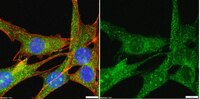
Polycystin-1 binds Par3/aPKC and controls convergent extension during renal tubular morphogenesis.
Castelli, M; Boca, M; Chiaravalli, M; Ramalingam, H; Rowe, I; Distefano, G; Carroll, T; Boletta, A
Nature communications
4
2658
2013
Show Abstract
Several organs, including the lungs and kidneys, are formed by epithelial tubes whose proper morphogenesis ensures correct function. This is best exemplified by the kidney, where defective establishment or maintenance of tubular diameter results in polycystic kidney disease, a common genetic disorder. Most polycystic kidney disease cases result from loss-of-function mutations in the PKD1 gene, encoding Polycystin-1, a large receptor of unknown function. Here we demonstrate that PC-1 has an essential role in the establishment of correct tubular diameter during nephron development. Polycystin-1 associates with Par3 favouring the assembly of a pro-polarizing Par3/aPKC complex and it regulates a programme of cell polarity important for oriented cell migration and for a convergent extension-like process during tubular morphogenesis. Par3 inactivation in the developing kidney results in defective convergent extension and tubular morphogenesis, and in renal cyst formation. Our data define Polycystin-1 as central to cell polarization and to epithelial tube morphogenesis and homeostasis. | Western Blotting, Immunofluorescence | | 24153433
|
Par-complex aPKC and Par3 cross-talk with innate immunity NF-κB pathway in epithelial cells.
Forteza, R; Wald, FA; Mashukova, A; Kozhekbaeva, Z; Salas, PJ
Biology open
2
1264-9
2013
Show Abstract
Components of the Par-complex, atypical PKC and Par3, have been found to be downregulated upon activation of NF-κB in intestinal epithelial cells. To determine their possible role in pro-inflammatory responses we transduced Caco-2 human colon carcinoma cells with constitutively active (ca) PKCι or anti-Par3 shRNA-expressing lentiviral particles. Contrary to previous reports in other cell types, ca-PKCι did not activate, but rather decreased, baseline NF-κB activity in a luminiscence reporter assay. An identical observation applied to a PB1 domain deletion PKCι, which fails to localize to the tight-junction. Conversely, as expected, the same ca-PKCι activated NF-κB in non-polarized HEK293 cells. Likewise, knockdown of Par3 increased NF-κB activity and, surprisingly, greatly enhanced its response to TNFα, as shown by transcription of IL-8, GRO-1, GRO-2 and GRO-3. We conclude that aPKC and Par3 are inhibitors of the canonical NF-κB activation pathway, although perhaps acting through independent pathways, and may be involved in pro-inflammatory responses. | | | 24244864
|


[07-330_WB_Final-2 (1)-ALL].jpg)
