Millipore Sigma Vibrant Logo

MABC1613 Anti-MUC1 Antibody, clone HMFG2

View Products on Sigmaaldrich.com
MABC1613
100 μg  
Prix en cours de récupération
Le prix n'a pas pu être récupéré
La quantité minimale doit être un multiple de
Maximum Quantity is
À la validation de la commande Plus d'informations
Vous avez sauvegardé ()
 
Demander le prix
Disponibilité limitée
En stock 
Interrompu(e)
Disponible en quantités limitées
Disponibilité à confirmer
    Pour le restant : Nous vous tiendrons informé
      Pour le restant : Nous vous tiendrons informé
      Nous vous tiendrons informé
      Contacter le Service Clients
      Contact Customer Service


      Contacter le Service Clients

      Aperçu

      Replacement Information

      Tableau de caractéristiques principal

      Species ReactivityKey ApplicationsHostFormatAntibody Type
      HIP, IHC, FUNC, WB, IH(P)MPurifiedMonoclonal Antibody
      Description
      Catalogue NumberMABC1613
      DescriptionAnti-MUC1 Antibody, clone HMFG2
      Alternate Names
      • Mucin-1
      • Breast carcinoma-associated antigen DF3
      • Cancer antigen 15-3
      • CA 15-3
      • Carcinoma-associated mucin
      • Episialin
      • H23AG
      • Krebs von den Lungen-6
      • KL-6
      • PEMT
      • Peanut-reactive urinary mucin
      • PUM
      • Polymorphic epithelial mucin
      • PEM
      • Tumor-associated epithelial membrane antigen
      • EMA
      • Tumor-associated mucin
      • CD227
      Background InformationMucin-1 (UniProt: P15941; MUC-1, Breast carcinoma-associated antigen DF3, Cancer antigen 15-3, CA 15-3, Carcinoma-associated mucin, Episialin, H23AG, Krebs von den Lungen-6, KL-6, PEMT, Peanut-reactive urinary mucin, PUM, Polymorphic epithelial mucin, PEM, Tumor-associated epithelial membrane antigen, EMA, Tumor-associated mucin, CD227) is encoded by the MUC1 (also known as PUM) gene (gene ID: 4582) in human. The MUC-1 alpha subunit has cell adhesive properties and functions as protective layer on epithelial cells against bacteria and enzymes. It can act as both cell adhesion and an anti-adhesion protein. It forms a tight, non-covalent heterodimeric complex with the proteolytically-released beta-subunit. The beta subunit contains a C-terminal domain that is involved in cell signaling via phosphorylations and protein-protein interactions. It modulates signaling in ERK, SRC and NF-kappa-B pathways. In activated T-cells, it impacts directly or indirectly the Ras/MAPK pathway. MUC-1 also is involved in tumor progression and transcription through regulation of Tp53. MUC-1 is expressed in epithelial cells, breast, uterus, activated and unactivated T-cells. It is shown to be over-expressed in epithelial tumors, such as breast or ovarian cancer. MUC-1 is exclusively located in the apical domain of the plasma membrane of highly polarized epithelial cells. After endocytosis, it is internalized and recycled to the cell membrane. MUC-1 is highly glycosylated (N- and O-linked carbohydrates and sialic acid) and is O-glycosylated to a varying degree on serine and threonine residues within each tandem repeat, ranging from mono- to penta-glycosylation. The average density ranges from about 50% in human milk to over 90% in T47D breast cancer cells. Its interaction, via the tandem repeat region, with domain 1 of ICAM1 is implicated in cell migration and metastases. MUC1 serves as a serological clinical marker of breast cancer to monitor response to breast cancer treatment and disease recurrence. Lower levels over time may be indicative of a positive response to treatment.
      References
      Product Information
      FormatPurified
      PresentationPurified mouse monoclonal antibody IgG1 in buffer containing 0.1 M Tris-Glycine (pH 7.4), 150 mM NaCl with 0.05% sodium azide.
      Quality LevelMQ100
      Applications
      Application
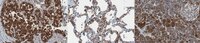
Detect Mucin-1 using this mouse monoclonal Anti-MUC1, clone HMFG2, Cat. No. MABC1613 that has been tested for use in Immunohistochemistry, Immunohistochemistry (Paraffin), Immunoprecipitation, Functional studies, and Western Blotting.
      Key Applications
      • Immunoprecipitation
      • Immunohistochemistry
      • Affects Function
      • Western Blotting
      • Immunohistochemistry (Paraffin)
      Application NotesImmunohistochemistry Analysis: A 1:1,000 dilution from a representative lot detected MUC1 in human lung and human pancreas tissue.

      Western Blotting Analysis: A representative lot detected MUC1 in Western Blotting applications (Kaur, S., et. al. (2014). PLoS One. 9(3):e92742; Schroeder, J.A., et. al. (2001). J Biol Chem. 276(16):13057-64).

      Affects Function: A representative lot detected MUC1 in Affects Function applications (Schettini, J., et. al. (2012). Cancer Immunol Immunother. 61(11):2055-65).

      Immunoprecipitation Analysis: A representative lot detected MUC1 in Immunoprecipitation applications in (Schroeder, J.A., et. al. (2001). J Biol Chem. 276(16):13057-64).

      Immunohistochemistry Analysis: A representative lot detected MUC1 in Immunohistochemistry applications in (Sakurai, J., et. al. (2007). Eur J Histochem. 51(2):95-102).
      Biological Information
      ImmunogenDelipidated milk fat globule extract followed 3 weeks later by epithelial cells cultured from milk (for 2 weeks).
      CloneHMFG2
      ConcentrationPlease refer to lot specific datasheet.
      HostMouse
      SpecificityClone HMFG2 immunostains human breast cancer tissue. It can react with the unglycosylated peptide as well as several glycoforms of MUC1.
      IsotypeIgG1λ
      Species Reactivity
      • Human
      Species Reactivity NoteHuman.
      Antibody TypeMonoclonal Antibody
      Entrez Gene Number
      Gene Symbol
      • MUC1
      • PUM
      Purification MethodProtein G purified
      UniProt Number
      Molecular Weight122.10 kDa calculated.
      Physicochemical Information
      Dimensions
      Materials Information
      Toxicological Information
      Safety Information according to GHS
      Safety Information
      Product Usage Statements
      Quality AssuranceEvaluated by Immunohistochemistry in human breast cancer tissue.

      Immunohistochemistry Analysis: A 1:1,000 dilution of this antibody detected MUC1 in human breast cancer tissue.
      Usage Statement
      • Unless otherwise stated in our catalog or other company documentation accompanying the product(s), our products are intended for research use only and are not to be used for any other purpose, which includes but is not limited to, unauthorized commercial uses, in vitro diagnostic uses, ex vivo or in vivo therapeutic uses or any type of consumption or application to humans or animals.
      Storage and Shipping Information
      Storage ConditionsStable for 1 year at 2-8°C from date of receipt.
      Packaging Information
      Material Size100 μg
      Transport Information
      Supplemental Information
      Specifications
      Global Trade Item Number
      Référence GTIN
      MABC1613 04054839197840