Millipore Sigma Vibrant Logo

AP1140 Anti-GBA Mouse mAb (2E2)

Aperçu

Replacement Information

Tableau de caractéristiques principal

Species ReactivityHostAntibody Type
HMMonoclonal Antibody

Prix & Disponibilité

Référence DisponibilitéConditionnement Qté Prix Quantité
AP1140-100UG
Récupération des données relatives à la disponibilité...
Disponibilité limitée
En stock 
Interrompu(e)
Disponible en quantités limitées
Disponibilité à confirmer
    Pour le restant : Nous vous tiendrons informé
      Pour le restant : Nous vous tiendrons informé
      Nous vous tiendrons informé
      Contacter le Service Clients
      Contact Customer Service

      100 μg
      Prix en cours de récupération
      Le prix n'a pas pu être récupéré
      La quantité minimale doit être un multiple de
      Maximum Quantity is
      À la validation de la commande Plus d'informations
      Vous avez sauvegardé ()
       
      Demander le prix
      Description
      OverviewRecognizes the ~60 kDa GBA protein in MCF-7 and HeLa cells and human breast cancer tissue.
      Catalogue NumberAP1140
      Brand Family Calbiochem®
      SynonymsAnti-Glucosidase β, Acid
      Application Data
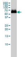
      Detection of human GBA by immunoblotting. Sample: Whole cell lysate from MCF-7 cells. Primary antibody: Anti-GBA Mouse mAb (2E2) (Cat. No. AP1140) (1-5 µg/ml). Detection: chemiluminescence.

      Detection of human GBA by immunocytochemistry. Sample: HeLa cells fixed in paraformaldehyde. Primary antibody: Anti-GBA Mouse mAb (2E2) (Cat. No. AP1140) (10 µg/ml). Detection: fluorescence.

      Detection of human GBA by staining paraffin sections. Sample: Formalin-fixed, paraffin-embedded human breast cancer tissue. Primary antibody: Anti-GBA Mouse mAb (2E2) (Cat. No. AP1140) (3 µg/ml). Detection: colorimetric (DAB).
      References
      ReferencesCampeau, P.M., et al. 2009. Blood 114, 3181.
      Product Information
      FormLiquid
      FormulationIn PBS, pH 7.2.
      Negative control293T
      Positive controlMCF-7 cells, HeLa cells, Human breast cancer tissue
      PreservativeNone
      Quality LevelMQ100
      Applications
      Key Applications Enzyme-Linked Immunosorbent Assay
      Immunoblotting (Western Blotting)
      Immunocytochemistry
      Paraffin Sections
      Application NotesELISA (see comments)
      Immunoblotting (1-5 µg/ml)
      Immunocytochemistry (10 µg/ml)
      Paraffin Sections (3 µg/ml)
      Application CommentsFor ELISA, this antibody can be used as the capture antibody; recommended concentration is 10 ng/ml, 1 µg/well, 100 µl total volume. Variables associated with assay conditions will dictate the proper working dilution.
      Biological Information
      ImmunogenA recombinant polypeptide corresponding to amino acids of 146-236 human GBA, expressed as a GST fusion protein
      Clone2E2
      HostMouse
      IsotypeIgG2a
      Species Reactivity
      • Human
      Antibody TypeMonoclonal Antibody
      Physicochemical Information
      Dimensions
      Materials Information
      Toxicological Information
      Safety Information according to GHS
      Safety Information
      Product Usage Statements
      Storage and Shipping Information
      Ship Code Blue Ice Only
      Toxicity Regulatory Review
      Storage -20°C
      Avoid freeze/thaw Avoid freeze/thaw
      Do not freeze Ok to freeze
      Special InstructionsFollowing initial thaw, aliquot and freeze (-20°C).
      Packaging Information
      Transport Information
      Supplemental Information
      Specifications
      Global Trade Item Number
      Référence GTIN
      AP1140-100UG 04055977227857

      Documentation

      Anti-GBA Mouse mAb (2E2) Certificats d'analyse

      TitreNuméro de lot
      AP1140

      Références bibliographiques

      Aperçu de la référence bibliographique
      Campeau, P.M., et al. 2009. Blood 114, 3181.
      Fiche technique

      Note that this data sheet is not lot-specific and is representative of the current specifications for this product. Please consult the vial label and the certificate of analysis for information on specific lots. Also note that shipping conditions may differ from storage conditions.

      Revision18-October-2010 RFH
      SynonymsAnti-Glucosidase β, Acid
      ApplicationELISA (see comments)
      Immunoblotting (1-5 µg/ml)
      Immunocytochemistry (10 µg/ml)
      Paraffin Sections (3 µg/ml)
      Application Data
      Detection of human GBA by immunoblotting. Sample: Whole cell lysate from MCF-7 cells. Primary antibody: Anti-GBA Mouse mAb (2E2) (Cat. No. AP1140) (1-5 µg/ml). Detection: chemiluminescence.

      Detection of human GBA by immunocytochemistry. Sample: HeLa cells fixed in paraformaldehyde. Primary antibody: Anti-GBA Mouse mAb (2E2) (Cat. No. AP1140) (10 µg/ml). Detection: fluorescence.

      Detection of human GBA by staining paraffin sections. Sample: Formalin-fixed, paraffin-embedded human breast cancer tissue. Primary antibody: Anti-GBA Mouse mAb (2E2) (Cat. No. AP1140) (3 µg/ml). Detection: colorimetric (DAB).
      DescriptionPurified mouse monoclonal antibody. Recognizes the ~60 kDa GBA protein.
      BackgroundAcid beta-glucosidase (GBA) is a lysosomal membrane protein that cleaves the beta-glucosidic linkage of glycosylceramide, an intermediate in glycolipid metabolism. It catalyzes the breakdown of the glycolipid glucosylceramide to ceramide and glucose. Mutations in this gene cause Gaucher disease, a lysosomal storage disease characterized by an accumulation of glucocerebrosides.
      HostMouse
      ImmunogenA recombinant polypeptide corresponding to amino acids of 146-236 human GBA, expressed as a GST fusion protein
      Clone2E2
      IsotypeIgG2a
      Specieshuman
      Positive controlMCF-7 cells, HeLa cells, Human breast cancer tissue
      Negative control293T
      FormLiquid
      FormulationIn PBS, pH 7.2.
      PreservativeNone
      CommentsFor ELISA, this antibody can be used as the capture antibody; recommended concentration is 10 ng/ml, 1 µg/well, 100 µl total volume. Variables associated with assay conditions will dictate the proper working dilution.
      Storage Avoid freeze/thaw
      -20°C
      Do Not Freeze Ok to freeze
      Special InstructionsFollowing initial thaw, aliquot and freeze (-20°C).
      Toxicity Regulatory Review
      ReferencesCampeau, P.M., et al. 2009. Blood 114, 3181.