Le fait de fermer ne sauvegardera pas votre configuration à moins que vous n'ajoutiez l'article à votre Panier d'achat ou à vos Favoris.
Cliquer sur OK pour fermer l'outil MILLIPLEX® MAP ou sur Annuler pour retourner à votre sélection.
Choisissez des Panels configurables & des Kits préconfigurés - OU - des MAPmate™ de signalisation cellulaire
Concevez vos kits MILLIPLEX® MAP et obtenez leur prix.
Panels configurables & Kits préconfigurés
Notre large gamme est constituée de panels multiplex qui vous permettent de choisir, au sein d'un panel, les analytes qui répondent le mieux à vos besoins. Sur un autre onglet, vous pouvez choisir un format cytokine préconfiguré ou un kit Simplex.
Kits de signalisation cellulaire & MAPmate™
Choisissez des kits préconfigurés qui permettent d'explorer l'ensemble des voies ou des processus. Ou concevez vos propres kits en choisissant des Simplex MAPmate™ et en suivant les instructions fournies.
Les MAPmate™ suivants ne peuvent pas être utilisés ensemble : -des MAPmate™ qui nécessitent des tampons différents -des paires de MAPmate™ totaux et phospho-spécifiques, par ex. GSK3β total et GSK3β (Ser 9) -des MAPmate™ PanTyr et spécifiques d'un site, par ex. Récepteur Phospho-EGF et phospho-STAT1 (Tyr701) -Plus d'un phospho-MAPmate™ pour une seule cible (Akt, STAT3). -GAPDH et β-Tubuline ne peuvent pas être utilisés avec les kits ou les MAPmate™ contenant panTyr.
.
Référence
Guide d'achat
Qté
Liste
Cet article a été ajouté à vos favoris.
Sélectionner une espèce, un type de panel, un kit ou un type d'échantillon
Pour commencer à concevoir votre kit MILLIPLEX® MAP, sélectionnez une espèce, un type de panel ou un kit d'intérêt.
Custom Premix Selecting "Custom Premix" option means that all of the beads you have chosen will be premixed in manufacturing before the kit is sent to you.
Catalogue Number
Ordering Description
Qty/Pack
List
Cet article a été ajouté à vos favoris.
Espèce
Type de panel
Kit sélectionné
Qté
Référence
Guide d'achat
Qté
Prix tarif
96-Well Plate
Qté
Référence
Guide d'achat
Qté
Prix tarif
Ajouter des réactifs supplémentaires (Un kit "Buffer and Detection Kit" est nécessaire pour une utilisation avec les MAPmate™)
Qté
Référence
Guide d'achat
Qté
Prix tarif
48-602MAG
Buffer Detection Kit for Magnetic Beads
1 Kit
Option de gain de place Nos clients qui commandent plusieurs kits peuvent choisir d'économiser de l'espace de stockage en éliminant l'emballage de chaque kit et de recevoir les composants de leur essai multiplex conditionnés sous poches en plastique pour un stockage plus compact.
Cet article a été ajouté à vos favoris.
Ce produit a été ajouté à votre panier.
Vous pouvez maintenant concevoir un autre kit personnalisé, choisir un kit pré-configuré, régler vos achats ou fermer l'outil de commande.
NA81
Sigma-AldrichAnti-5-Methylcytosine Mouse mAb (162 33 D3)
This Anti-5-Methylcytosine Mouse mAb (162 33 D3) is validated for use in ELISA, FC, Frozen Sections, IF, Paraffin Sections, Radioimmunoassay, Southwestern Blot for the detection of 5-Methylcytosine.
More>>This Anti-5-Methylcytosine Mouse mAb (162 33 D3) is validated for use in ELISA, FC, Frozen Sections, IF, Paraffin Sections, Radioimmunoassay, Southwestern Blot for the detection of 5-Methylcytosine. Less<<
Anti-5-Methylcytosine Mouse mAb (162 33 D3) : FDS (Fiches de données de sécurité), certificats d’analyse (CoA) et de qualité (CoQ), dossiers, brochures et autres documents disponibles.
Le champ quantité est vide. Veuillez saisir une quantité supérieure ou égale à 1 au minimum pour ajouter des articles à votre panier.
Description
Overview
Recognizes 5-methylcytosine in methylated DNA or RNA in NIH3T3 cells.
Specific • Affinity purified mouse monoclonal antibody - Detects methylated DNA from a broad range of species.
Several Applications • Flow Cytometry
• Frozen Sections
• Immunoblotting
• Immunofluorescence
• Paraffin Sections
Reliable • Generates reproducible results.
• Clone 162 33 D3 has been used in over 50 publications.
Catalogue Number
NA81
Brand Family
Calbiochem®
Synonyms
Anti-5-mc, Anti-5-MeCyd
Application Data
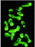
Detection of 5-methylcytosine by immunohistochemistry. Sample: Normal colon (left) and colon adenocarcinoma (right). Anti-5-Methylcytosine Mouse mAb (162 33 D3) (Cat. No. NA81). Detection: DAB Chromosome methylation patterns of mouse embryos at the one-cell stage. Methylated sites were revealed by indirect immunofluorescence labeling with Anti-5-methylcytosine.
References
References
Ehrlich, M. 2002. Oncogene21, 5400. (Review) Widschwendter, M. and Jones, P.A. 2002. Oncogene21, 5462. (Review) Barton, S.C., et al. 2001. Hum. Mol. Genet.10, 2983. Piyathilake, C.J., et al. 2001. Hum. Pathol.32, 856. Taddei, A., et al. 2001. Nat. Cell Biol.3, 114. Piyathilake, C.J., et al. 2000. Biotech. Histochem.75, 251. De Capoa, A., et al. 1999. FASEB J.13, 89.
Clone 162 33 D3 is useful for the quantitative and qualitative detection of methylated DNA or RNA in a variety of samples and applications. It has been used in over 40 publications. Antibody should be titrated for optimal results in an individual systems.
Biological Information
Immunogen
5-methylcytosine conjugated to ovalbumin
Clone
162 33 D3
Host
Mouse
Isotype
IgG₁
Species Reactivity
A Broad Range Of Species
Antibody Type
Monoclonal Antibody
Concentration Label
Please refer to vial label for lot-specific concentration
Storage and Shipping Information
Ship Code
Blue Ice Only
Toxicity
Standard Handling
Storage
-20°C
Avoid freeze/thaw
Avoid freeze/thaw
Do not freeze
Ok to freeze
Special Instructions
Following initial thaw, aliquot and freeze (-20°C).
Note that this data sheet is not lot-specific and is representative of the current specifications for this product. Please consult the vial label and the certificate of analysis for information on specific lots. Also note that shipping conditions may differ from storage conditions.
Detection of 5-methylcytosine by immunohistochemistry. Sample: Normal colon (left) and colon adenocarcinoma (right). Anti-5-Methylcytosine Mouse mAb (162 33 D3) (Cat. No. NA81). Detection: DAB Chromosome methylation patterns of mouse embryos at the one-cell stage. Methylated sites were revealed by indirect immunofluorescence labeling with Anti-5-methylcytosine.
DNA methylation plays an important role in gene regulation. Methylation of gene-promoter regions leads to loss of function while DNA demethylation can lead to gain of function. Alterations in DNA methylation may play an important role in carcinogenesis. DNA methylation varies according to tissue type. In some cancers types such as Wilms tumor and colon adenocarcinoma have a decrease in global DNA methylation. In contrast other cancers such as breast cancer have DNA hypermethylation. Changes in global DNA methylation status may be useful early markers for the detection of premalignant lesions.
Host
Mouse
Immunogen
5-methylcytosine conjugated to ovalbumin
Clone
162 33 D3
Isotype
IgG₁
Species
a broad range of species
Positive control
NIH3T3 cells
Form
Liquid
Formulation
In PBS, pH 7.4.
Concentration Label
Please refer to vial label for lot-specific concentration
Preservative
None
Comments
Clone 162 33 D3 is useful for the quantitative and qualitative detection of methylated DNA or RNA in a variety of samples and applications. It has been used in over 40 publications. Antibody should be titrated for optimal results in an individual systems.
Storage
Avoid freeze/thaw
-20°C
Do Not Freeze
Ok to freeze
Special Instructions
Following initial thaw, aliquot and freeze (-20°C).
Toxicity
Standard Handling
References
Ehrlich, M. 2002. Oncogene21, 5400. (Review) Widschwendter, M. and Jones, P.A. 2002. Oncogene21, 5462. (Review) Barton, S.C., et al. 2001. Hum. Mol. Genet.10, 2983. Piyathilake, C.J., et al. 2001. Hum. Pathol.32, 856. Taddei, A., et al. 2001. Nat. Cell Biol.3, 114. Piyathilake, C.J., et al. 2000. Biotech. Histochem.75, 251. De Capoa, A., et al. 1999. FASEB J.13, 89.
Application references
Paraffin Sections
Kang, J.S., et al. 2006. Cancer Sci.97, 453.