


NR07 Sigma-AldrichAnti-Ryanodine Receptor Mouse mAb (C3-33)

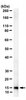
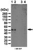
This Anti-Ryanodine Receptor Mouse mAb (C3-33) is validated for use in Frozen Sections, Immunoblotting, Immunoprecipitation for the detection of Ryanodine Receptor.
More>> This Anti-Ryanodine Receptor Mouse mAb (C3-33) is validated for use in Frozen Sections, Immunoblotting, Immunoprecipitation for the detection of Ryanodine Receptor. Less<<Anti-Ryanodine Receptor Mouse mAb (C3-33) MSDS (material safety data sheet) or SDS, CoA and CoQ, dossiers, brochures and other available documents.
Recommended Products


Overview
| Replacement Information |
|---|
Key Spec Table
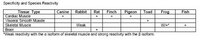
| Species Reactivity | Host | Antibody Type |
|---|---|---|
| Am, Av, Ca, F, Rb, R | M | Monoclonal Antibody |
Pricing & Availability
| Catalogue Number | Availability | Packaging | Qty/Pack | Price | Quantity | |
|---|---|---|---|---|---|---|
| NR07-100UG |
|
Plastic ampoule | 100 μg |
|
— |


| Product Information | |
|---|---|
| Form | Liquid |
| Formulation | In PBS. |
| Positive control | Rat brain hippocampus |
| Preservative | ≤0.1% sodium azide |
| Quality Level | MQ100 |
| Physicochemical Information |
|---|
| Dimensions |
|---|
| Materials Information |
|---|
| Toxicological Information |
|---|
| Safety Information according to GHS |
|---|
| Safety Information |
|---|
| Product Usage Statements |
|---|
| Packaging Information |
|---|
| Transport Information |
|---|
| Supplemental Information |
|---|
| Specifications |
|---|
| Global Trade Item Number | |
|---|---|
| Catalogue Number | GTIN |
| NR07-100UG | 04055977210002 |
Documentation
Anti-Ryanodine Receptor Mouse mAb (C3-33) SDS
| Title |
|---|
Anti-Ryanodine Receptor Mouse mAb (C3-33) Certificates of Analysis
| Title | Lot Number |
|---|---|
| NR07 |
References
| Reference overview |
|---|
| Sitsapesan, R., et al. 1995. Trends Pharmacol. Sci. 16, 386. McPherson, P.S. and Campbell, K.P. 1993. J. Biol. Chem. 268, 13765. Meszaros, L.G., et al. 1993. Nature 364, 76. Lai, F.A., et al. 1992. Biochem. J. 288, 553. Lai, F.A., et al. 1989. Nature 331, 315. |