Millipore Sigma Vibrant Logo

MABE1110 Anti-PASD1 Antibody, clone 2ALCC136 (PASD1-1)

View Products on Sigmaaldrich.com
MABE1110
100 μg  
Retrieving price...
Price could not be retrieved
Minimum Quantity is a multiple of
Maximum Quantity is
Upon Order Completion More Information
You Saved ()
 
Request Pricing
Fulfillment and Delivery Delayed
In Stock 
Discontinued
Limited Quantities Available
Availability to be confirmed
    Remaining : Will advise
      Remaining : Will advise
      Will advise
      Contact Customer Service
      Contact Customer Service

      Special Offers


      Contact Customer Service

      Overview

      Replacement Information

      Key Spec Table

      Species ReactivityKey ApplicationsHostFormatAntibody Type
      HICC, IH(P), WBMPurifiedMonoclonal Antibody
      Description
      Catalogue NumberMABE1110
      DescriptionAnti-PASD1 Antibody, clone 2ALCC136 (PASD1-1)
      Alternate Names
      • Circadian clock protein PASD1
      • Cancer/testis antigen 63
      • CT63
      • OX-TES-1
      • PAS domain-containing protein 1
      Background InformationCircadian clock protein PASD1 (UniProt Q8IV76; also known as Cancer/testis antigen 63, CT63, OX-TES-1, PAS domain-containing protein 1) is encoded by the PASD1 gene (Gene ID 139135) in human. Originally characterized as a lymphoma-associated antigen and a candidate cancer/testis antigen (CTA), PASD1 is evolutionarily related to CLOCK and interacts with the bHLH-PAS transcription complex CLOCK-BMAL1 to repress transcriptional activation. PASD1 expression is normally limited to immunologically restricted germline tissues that do not express class I MHC molecules, such as testis and placenta, allowing tissue-specific circadian rhythms suppression. PASD1 can be induced in cells of somatic origin upon oncogenic transformation and PASD1 is among the most frequently expressed acute myeloid leukemia (AML) tumour antigens. Reducing PASD1 in human cancer cells significantly increases the amplitude of transcriptional oscillations to generate more robust circadian rhythms. Human PASD1 gene is located on Xq28, and mutations in the q28 region have been linked with increased risk of lymphoma and leukemia, Human PASD1 contains an N-terminal PAS domain (a.a. 30-102), a central region (a.a. 365-412) essential for its transcriptional repression activity, and two coiled coil domains (CC1/a.a. 365-412 and CC2/a.a. 475-553), where CC1 exhibits significant homology with CLOCK exon 19-coded sequence.
      References
      Product Information
      FormatPurified
      PresentationPurified mouse IgG2a in buffer containing 0.1 M Tris-Glycine (pH 7.4), 150 mM NaCl with 0.05% sodium azide.
      Quality LevelMQ100
      Applications
      ApplicationDetect PASD1 using this mouse monoclonal Anti-PASD1 Antibody, clone 2ALCC136 (PASD1-1), Cat. No. MABE1110, validated for use in Immunocytochemistry, Immunohistochemistry (Paraffin), and Western Blotting.
      Key Applications
      • Immunocytochemistry
      • Immunohistochemistry (Paraffin)
      • Western Blotting
      Application NotesImmunocytochemistry Analysis: Clone 2ALCC136 hybridoma culture supernatant detected PASD1 immunoreactivity in 3 out of 18 leukemia patient samples (Brooks, S.E., et al. (2015). PLoS One. 10(10):e0140483).

      Immunocytochemistry Analysis: Clone 2ALCC136 hybridoma culture supernatant immunostained chronic myeloid leukemia line K562 and multiple myeloma line THIEL (Khan, G., et al. (2015). Biomark Cancer. 7:31-38).

      Immunocytochemistry Analysis: Clone 2ALCC136 hybridoma culture supernatant detected differential PASD1 cellular localization among cancer cell lines, including Breast cancer (BT-20), colon cancer (SW480), lymphoma (Granta519, KM-H2, OCI-Ly3 DLBCL), NSCLC (COR-L23, NCI-H1299) lines (Michael, A.K., et al. (2015). Mol. Cell. 58(5):743-754; Cooper, C.D., et al. (2006). Leukemia. 20(12):2172-2174).

      Immunohistochemistry Analysis: Clone 2ALCC136 hybridoma culture supernatant determined PASD1 immunoreactivity in skin tumor samples, as well as ovarian cancer tissues of different disease stage (Khan, G., et al. (2015). Biomark Cancer. 7:31-38).

      Immunohistochemistry Analysis: Clone 2ALCC136 hybridoma culture supernatant immunostained spermatogonia nucleus in paraffin-embedded human testis sections, while predominant cytoplasmic staining of cancer cells was seen among lymphoma biopsies (Ait-Tahar, K., et al. (2009). Br. J. Haematol. 146(4):396-407; Cooper, C.D., et al. (2006). Leukemia. 20(12):2172-2174).

      Western Blotting Analysis: Clone 2ALCC136 hybridoma culture supernatant detected different PASD1 expression levels among a panel of human cancer cell lines, including melanoma, NSCLC, breast and colon cancers (Michael, A.K., et al. (2015). Mol. Cell. 58(5):743-754).

      Western Blotting Analysis: Clone 2ALCC136 hybridoma culture supernatant detected both endogenous (Thiel and KM-H2) and exogenously expressed (COS) ~100 kDa PASD1 long isoform, as well as a ~40 kDa isoform in OCI-Ly3 and FEPD cells (Cooper, C.D., et al. (2006). Leukemia. 20(12):2172-2174).
      Biological Information
      ImmunogenGST-tagged recombinant human PASD1 fragment corresponding to the region between the PAS domain and the second coiled coil region.
      Clone2ALCC136
      ConcentrationPlease refer to lot specific datasheet.
      HostMouse
      SpecificityClone 2ALCC136 (a.k.a. PASD1-1) recognizes both human PASD1 spliced isoforms reported by UniProt (Q8IV76), as well as a novel ~40 kDa isoform (Cooper, C.D., et al. (2006). Leukemia. 20(12):2172-2174).
      IsotypeIgG2aκ
      Species Reactivity
      • Human
      Species Reactivity NoteHuman.
      Antibody TypeMonoclonal Antibody
      Entrez Gene Number
      Gene Symbol
      • PASD1
      Purification MethodProtein G purified.
      UniProt Number
      Molecular Weight87.43 kDa (isoform 1) and 72.74 kDa (isoform 2) calculated. ~100 kDa (isoform 1) and ~40 kDa (a novel isoform) reported (Cooper, C.D., et al. (2006). Leukemia. 20(12):2172-2174). Note that Cooper, C.D., et al. (2006) referred to the long isoform (UniProt isoform 1) as PASD1_v1 and the short isoform (UniProt isoform 2) as PASD1_v2, while the exact opposite is assigned by UniProt (Q8IV76).
      Physicochemical Information
      Dimensions
      Materials Information
      Toxicological Information
      Safety Information according to GHS
      Safety Information
      Product Usage Statements
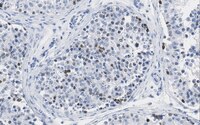
      Quality AssuranceEvaluated by Immunohistochemistry in human testis tissue sections.

      Immunhistochemistry Analysis: A 1:50 dilution of this antibody detected nuclear PASD1 immunoreactivity in human testis tissue section.
      Usage Statement
      • Unless otherwise stated in our catalog or other company documentation accompanying the product(s), our products are intended for research use only and are not to be used for any other purpose, which includes but is not limited to, unauthorized commercial uses, in vitro diagnostic uses, ex vivo or in vivo therapeutic uses or any type of consumption or application to humans or animals.
      Storage and Shipping Information
      Storage ConditionsStable for 1 year at 2-8°C from date of receipt.
      Packaging Information
      Material Size100 μg
      Transport Information
      Supplemental Information
      Specifications
      Global Trade Item Number
      Catalogue Number GTIN
      MABE1110 04054839078743

      Documentation

      Anti-PASD1 Antibody, clone 2ALCC136 (PASD1-1) SDS

      Title

      Safety Data Sheet (SDS) 

      Anti-PASD1 Antibody, clone 2ALCC136 (PASD1-1) Certificates of Analysis

      TitleLot Number
      Anti-PASD1, clone 2ALCC136 (PASD1-1) -Q2766922 Q2766922

      References

      Reference overviewPub Med ID
      Identification of survivin as a promising target for the immunotherapy of adult B-cell acute lymphoblastic leukemia.
      Boullosa LF, Savaliya P, Bonney S, Orchard L, Wickenden H, Lee C, Smits E, Banham AH, Mills KI, Orchard K, Guinn BA
      Oncotarget. 9(3):3853-3866 (2017)  2017

      Show Abstract
      29423088 29423088