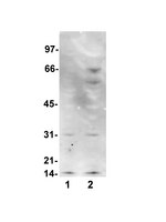
16-227 Sigma-AldrichAnti-Nitrotyrosine Antibody, clone 1A6, Alexa Fluor® 555 conjugate
Anti-Nitrotyrosine Antibody, clone 1A6, Alexa Fluor 555 conjugate is an antibody against Nitrotyrosine for use in FC & WB.
More>> Anti-Nitrotyrosine Antibody, clone 1A6, Alexa Fluor 555 conjugate is an antibody against Nitrotyrosine for use in FC & WB. Less<<Recommended Products
Overview
| Replacement Information |
|---|
Key Spec Table
| Species Reactivity | Key Applications | Host | Format | Antibody Type |
|---|---|---|---|---|
| Vrt | FC, WB | M | AlexaFluor®555 | Monoclonal Antibody |
| Description | |
|---|---|
| Catalogue Number | 16-227 |
| Brand Family | Upstate |
| Trade Name |
|
| Description | Anti-Nitrotyrosine Antibody, clone 1A6, Alexa Fluor® 555 conjugate |
| References |
|---|
| Product Information | |
|---|---|
| Format | AlexaFluor®555 |
| Quality Level | MQ100 |
| Applications | |
|---|---|
| Application | Anti-Nitrotyrosine Antibody, clone 1A6, Alexa Fluor 555 conjugate is an antibody against Nitrotyrosine for use in FC & WB. |
| Key Applications |
|
| Physicochemical Information |
|---|
| Dimensions |
|---|
| Materials Information |
|---|
| Toxicological Information |
|---|
| Safety Information according to GHS |
|---|
| Safety Information |
|---|
| Storage and Shipping Information | |
|---|---|
| Storage Conditions | 1 year at 4°C from date of shipment |
| Packaging Information | |
|---|---|
| Material Size | 100 µg |
| Transport Information |
|---|
| Supplemental Information |
|---|
| Specifications |
|---|
| Global Trade Item Number | |
|---|---|
| Catalogue Number | GTIN |
| 16-227 | 04053252268892 |
Documentation
Anti-Nitrotyrosine Antibody, clone 1A6, Alexa Fluor® 555 conjugate Certificates of Analysis
References
| Reference overview | Application | Pub Med ID |
|---|---|---|
| Biological significance of nitric oxide-mediated protein modifications. Gow, Andrew J, et al. Am. J. Physiol. Lung Cell Mol. Physiol., 287: L262-8 (2004) 2004 Show Abstract | 15246980
 | |
| Nitric oxide (NO) induces nitration of protein kinase Cepsilon (PKCepsilon ), facilitating PKCepsilon translocation via enhanced PKCepsilon -RACK2 interactions: a novel mechanism of no-triggered activation of PKCepsilon Balafanova, Z., et al J Biol Chem, 277:15021-7 (2002) 2002 | Immunoblotting (Western), Immunoprecipitation | 11839754
 |
| Oxidation of the zinc-thiolate complex and uncoupling of endothelial nitric oxide synthase by peroxynitrite Zou, M. H., et al J Clin Invest, 109:817-26 (2002) 2002 | Immunoblotting (Western) | 11901190
 |
| Reactive oxygen nitrogen species decrease cystic fibrosis transmembrane conductance regulator expression and cAMP-mediated Cl- secretion in airway epithelia♪A mutation in the cystic fibrosis transmembrane conductance regulator generates a novel internali Bebok, Z., et al J Biol Chem, 277:43041-9 (2002) 2002 | Immunoblotting (Western) | 12167629
 |
| In vitro production of peroxynitrite by haemocytes from marine bivalves: C-ELISA determination of 3-nitrotyrosine level in plasma proteins from Mytilus galloprovincialis and Crassostrea gigas Torreilles, J. and Romestand, B. BMC Immunol, 2:1 (2001) 2001 | Enzyme Immunoassay (ELISA) | 11231884
 |
| Immunohistochemical methods to detect nitrotyrosine Viera, L., et al Methods Enzymol, 301:373-81 (1999) 1999 | Immunohistochemistry (Tissue) | 9919586
 |
| Mechanisms of reduced striatal NMDA excitotoxicity in type I nitric oxide synthase knock-out mice. Ayata, C, et al. J. Neurosci., 17: 6908-17 (1997) 1997 Show Abstract | 9278526
 | |
| Nitration and inactivation of manganese superoxide dismutase in chronic rejection of human renal allografts. MacMillan-Crow, L A, et al. Proc. Natl. Acad. Sci. U.S.A., 93: 11853-8 (1996) 1996 Show Abstract | 8876227
 | |
| Evidence for in vivo peroxynitrite production in human acute lung injury Kooy, N. W., et al Am J Respir Crit Care Med, 151:1250-4 (1995) 1995 | Immunohistochemistry (Tissue) | 7697261
 |
| Extensive nitration of protein tyrosines in human atherosclerosis detected by immunohistochemistry. Beckmann, J S, et al. Biol. Chem. Hoppe-Seyler, 375: 81-8 (1994) 1994 | 8192861
 |
Brochure
| Title |
|---|
| Pathways and Biomarkers of Oxidative Stress |
Technical Info
| Title |
|---|
| White Paper- Modern Methods in Oxidative Stress Research |


