Millipore Sigma Vibrant Logo

MABS1271 Anti-EPCR Antibody, clone JRK1535

View Products on Sigmaaldrich.com
MABS1271
100 μg  
Retrieving price...
Price could not be retrieved
Minimum Quantity is a multiple of
Maximum Quantity is
Upon Order Completion More Information
You Saved ()
 
Request Pricing
Fulfillment and Delivery Delayed
In Stock 
Discontinued
Limited Quantities Available
Availability to be confirmed
    Remaining : Will advise
      Remaining : Will advise
      Will advise
      Contact Customer Service
      Contact Customer Service

      Special Offers


      Contact Customer Service

      Overview

      Replacement Information

      Key Spec Table

      Species ReactivityKey ApplicationsHostFormatAntibody Type
      HFC, NEUTMPurifiedMonoclonal Antibody
      Description
      Catalogue NumberMABS1271
      DescriptionAnti-EPCR Antibody, clone JRK1535
      Alternate Names
      • Endothelial protein C receptor
      • Activated protein C receptor
      • APC receptor
      • CD201
      • Endothelial cell protein C receptor
      Background InformationEndothelial protein C receptor (UniProt Q9UNN8; also known as Activated protein C receptor, APC receptor, CD201, Endothelial cell protein C receptor) is encoded by the PROCR (also known as EPCR) gene (Gene ID 10544) in human. The coagulation cascade is an evolutionary conserved pathway in vertebrates that maintains vascular integrity. The procoagulant activity of the tssue factor (TF) pathway is counterbalanced by the protein C (PC) anticoagulant pathway to avoid intravascular thrombosis. EPCR is a CD1d–like immune receptor that binds the γ-carboxyl glutamic acid-rich (Gla) domain of PC and markedly improves PC activation in response to thrombin binding to endothelial cell-expressed thrombomodulin. EPCR also serves as the co-receptor for activated PC (aPC) in vascular protective signaling mediated by activation of PAR1. In addition, EPCR is found on hematopoietic, neuronal and epithelial progenitor populations, as well as among highly aggressive basal-like breast cancer subtypes. Endothelial overexpression of EPCR attenuates metastasis, presumably by dampening thrombin generation that supports metastatic tumor cell survival. EPCR-dependent PAR1 activation by aPC also stimulates breast cancer cell migration and prevents apoptosis of lung cancer cells. EPCR-selected populations of breast cancer cells grow as non-adherent spheroids and have high tumorigenicity when injected at low cell numbers. EPCR is initially produced with a signal peptide (a.a. 1-17), the removal of which yields the mature protein with a large extracellular region (a.a. 18-210), followed by a transmembrane domain (a.a. 211-231) and a short cytoplasmic tail (a.a. 232-238).
      References
      Product Information
      FormatPurified
      PresentationPurified mouse monoclonal IgG1κ antibody in PBS without preservatives.
      Quality LevelMQ100
      Applications
      ApplicationAnti-EPCR Antibody, clone JRK1535 is an antibody against EPCR for use in Flow Cytometry, Neutralizing.
      Key Applications
      • Flow Cytometry
      • Neutralizing
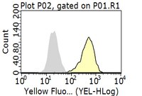
      Application NotesFlow Cytometry Analysis: 0.1 µg of this antibody from a representative lot detected EPCR in one million HeLa cells.
      Flow Cytometry Analysis: A representative lot, when conjugated with fluorescein, detected Doxorubicin (Cat. No. 324380) treatment-induced downregulation of EPCR expression on the surface of HUVECs. Glutathione pretreatment prevented Doxorubicin-induced EPCR downregulation (Woodley-Cook, J., et al. (2006). Mol. Cancer Ther. 5(12):3303-3311).
      Flow Cytometry Analysis: A representative lot blocked fluorescein-labeled APC from binding exogenously expressed human EPCR on the surface of HEK293 transfectants. When labeled with fluorescein, clone JRK1535 stained HEK293 transfectants expressing human EPCR, but not transfectants expressing murine EPCR (Liaw, P.C., et al. (2001). J. Biol. Chem. 276(11):8364-8370).
      Neutralizing Analysis: A representative lot inhibited APC generation from acrolein-treated HUVECs exposed to defibrinated, recalcified plasma (Swystun, L.L., et al. (2011). J Thromb Haemost. 9(4):767-775).
      Neutralizing Analysis: A representative lot selectively affected the survival of sorted EPCR-positive, but not EPCR-negative, MDA-MB-231 mammary fat pad (mfp) cells in cultures. Pretreatment of EPCR-positive cells with clone JRK1535 prior to xenografting also suppressed the tumor-initiating capacity of the EPCR-positive cells in mice vivo (Schaffner, F., et al. (2013). PLoS One. 8(4):e61071).
      Biological Information
      ImmunogenRecombinant human EPCR extracellular domain.
      EpitopeExtracellular domain.
      CloneJRK1535
      ConcentrationPlease refer to lot specific datasheet.
      HostMouse
      SpecificityClone JRK1535 blocked activated protein C (APC) from binding cell surface EPCR by targeting a conformational epitope at the extracellular domain (Liaw, P.C., et al. (2001). J. Biol. Chem. 276(11):8364-8370).
      IsotypeIgG1κ
      Species Reactivity
      • Human
      Species Reactivity NoteHuman. Not mouse.
      Antibody TypeMonoclonal Antibody
      Entrez Gene Number
      Gene Symbol
      • PROCR
      • EPCR
      Purification MethodProtein G Purified
      UniProt Number
      Molecular Weight24.80 kDa calculated. Greater than 40 kDa observed due to glycosylation (Woodley-Cook, J., et al. (2006). Mol. Cancer Ther. 5(12):3303-3311).
      Physicochemical Information
      Dimensions
      Materials Information
      Toxicological Information
      Safety Information according to GHS
      Safety Information
      Product Usage Statements
      Quality AssuranceEvaluated by Flow Cytometry in A549 cells.

      Flow Cytometry Analysis: 0.1 µg of this antibody detected EPCR in one million A549 cells.
      Usage Statement
      • Unless otherwise stated in our catalog or other company documentation accompanying the product(s), our products are intended for research use only and are not to be used for any other purpose, which includes but is not limited to, unauthorized commercial uses, in vitro diagnostic uses, ex vivo or in vivo therapeutic uses or any type of consumption or application to humans or animals.
      Storage and Shipping Information
      Storage ConditionsStable for 1 year at -20°C from date of receipt.
      Handling Recommendations: Upon receipt and prior to removing the cap, centrifuge the vial and gently mix the solution. Aliquot into microcentrifuge tubes and store at -20°C. Avoid repeated freeze/thaw cycles, which may damage IgG and affect product performance.
      Packaging Information
      Material Size100 μg
      Transport Information
      Supplemental Information
      Specifications
      Global Trade Item Number
      Catalogue Number GTIN
      MABS1271 04055977333572