Millipore Sigma Vibrant Logo

MABS1154 Anti-Derlin-1 Antibody, clone 8G11.2

View Products on Sigmaaldrich.com
MABS1154
100 μg  
Retrieving price...
Price could not be retrieved
Minimum Quantity is a multiple of
Maximum Quantity is
Upon Order Completion More Information
You Saved ()
 
Request Pricing
Fulfillment and Delivery Delayed
In Stock 
Discontinued
Limited Quantities Available
Availability to be confirmed
    Remaining : Will advise
      Remaining : Will advise
      Will advise
      Contact Customer Service
      Contact Customer Service

      Special Offers


      Contact Customer Service

      Overview

      Replacement Information

      Key Spec Table

      Species ReactivityKey ApplicationsHostFormatAntibody Type
      H, MWB, ICCMPurifiedMonoclonal Antibody
      Description
      Catalogue NumberMABS1154
      DescriptionAnti-Derlin-1 Antibody, clone 8G11.2
      Alternate Names
      • Degradation in endoplasmic reticulum protein 1
      • Der1-like protein 1
      • Derlin-1
      • DERtrin-1
      Background InformationDerlin-1 (UniProt: Q9BUN8; also known as Degradation in endoplasmic reticulum protein 1, DERtrin-1, Der1-like protein 1) is encoded by the DERL1 (also known as DER1, UNQ243/PRO276) gene (Gene ID: 79139) in human. Derlin-1 is a multi-pass membrane protein that serves as an E3 ligase mediator that links target proteins to ubiquitin-mediated proteasomal degradation in the cytosol. It is a functional sub-unit of the ER-associated degradation (ERAD) that forms a retro-translocation channel in the ER membrane and functions in the recognition of misfolded membrane or transmembrane proteins and their subsequent degradation in the ubiquitin-proteasomal system. Before any ERAD proteins are processed by the cytoplasmic ubiquitin-proteasome system, they are translocated across the ER membrane through an AAA ATPase p97/VCP-driven process. Derlin-1 is also involved in endoplasmic reticulum stress-induced pre-emptive quality control, a mechanism that selectively attenuates the translocation of newly synthesized proteins into the endoplasmic reticulum and reroutes them to the cytosol for proteasomal degradation. Derlin-1 can form homo- and heterooligomers with Derlin-2 and -3, however, binding to Derlin-3 is shown to be DERL3 is lower. Derlin-1 contains three cytoplasmic domains (aa 2-22, 81-105, and 176-251), four transmembrane domains (aa 23-43, 60-80, 106-126, 155-175), and two lumenal domains (aa 44-59 and 127-154). (Ref.: You, H., et al. (2017). J. Cell Sci. 130(6); 1027-1036).
      References
      Product Information
      FormatPurified
      PresentationPurified mouse monoclonal IgG2bκ antibody in buffer containing 0.1 M Tris-Glycine (pH 7.4), 150 mM NaCl with 0.05% sodium azide.
      Quality LevelMQ100
      Applications
      ApplicationThis Anti-Derlin-1 Antibody, clone 8G11.2 is validated for use in Western Blotting, Immunocytochemistry for the detection of Derlin-1.
      Key Applications
      • Western Blotting
      • Immunocytochemistry
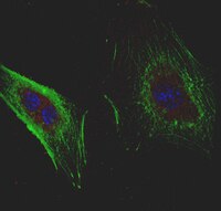
      Application NotesImmunocytochemistry Analysis: An 1:50 dilution from a representative lot detected Derlin-1 in HUVECs, A431 and HeLa cells.
      Biological Information
      ImmunogenGST-tagged recombinant human derlin-1 C-terminal cytoplasmic fragment.
      EpitopeC-terminal cytoplasmic domain.
      Clone8G11.2
      ConcentrationPlease refer to lot specific datasheet.
      HostMouse
      SpecificityExpected to react with both spliced isoforms of human Derlin-1 reported by UniProt (Q9BUN8).
      IsotypeIgG2bκ
      Species Reactivity
      • Human
      • Mouse
      Antibody TypeMonoclonal Antibody
      Entrez Gene Number
      Gene Symbol
      • DERL1
      • DER1
      • UNQ243/PRO276
      Purification MethodProtein G Purified
      UniProt Number
      Molecular Weight~24 kDa observed. 28.80 kDa (isoform 1) and 26.42 kDa (isoform 2) calculated.
      Physicochemical Information
      Dimensions
      Materials Information
      Toxicological Information
      Safety Information according to GHS
      Safety Information
      Product Usage Statements
      Quality AssuranceEvaluated by Western Blotting in HEK293 cell lysate.

      Western Blotting Analysis: 1.0 µg/mL of this antibody detected Derlin-1 in 10 µg of HEK293 cell lysate.
      Usage Statement
      • Unless otherwise stated in our catalog or other company documentation accompanying the product(s), our products are intended for research use only and are not to be used for any other purpose, which includes but is not limited to, unauthorized commercial uses, in vitro diagnostic uses, ex vivo or in vivo therapeutic uses or any type of consumption or application to humans or animals.
      Storage and Shipping Information
      Storage ConditionsStable for 1 year at 2-8°C from date of receipt.
      Packaging Information
      Material Size100 μg
      Transport Information
      Supplemental Information
      Specifications
      Global Trade Item Number
      Catalogue Number GTIN
      MABS1154 04055977331202