Wenn Sie das Fenster schließen, wird Ihre Konfiguration nicht gespeichert, es sei denn, Sie haben Ihren Artikel in die Bestellung aufgenommen oder zu Ihren Favoriten hinzugefügt.
Klicken Sie auf OK, um das MILLIPLEX® MAP-Tool zu schließen oder auf Abbrechen, um zu Ihrer Auswahl zurückzukehren.
Wählen Sie konfigurierbare Panels & Premixed-Kits - ODER - Kits für die zelluläre Signaltransduktion & MAPmates™
Konfigurieren Sie Ihre MILLIPLEX® MAP-Kits und lassen sich den Preis anzeigen.
Konfigurierbare Panels & Premixed-Kits
Unser breites Angebot enthält Multiplex-Panels, für die Sie die Analyten auswählen können, die am besten für Ihre Anwendung geeignet sind. Unter einem separaten Register können Sie das Premixed-Cytokin-Format oder ein Singleplex-Kit wählen.
Kits für die zelluläre Signaltransduktion & MAPmates™
Wählen Sie gebrauchsfertige Kits zur Erforschung gesamter Signalwege oder Prozesse. Oder konfigurieren Sie Ihre eigenen Kits mit Singleplex MAPmates™.
Die folgenden MAPmates™ sollten nicht zusammen analysiert werden: -MAPmates™, die einen unterschiedlichen Assaypuffer erfordern. -Phosphospezifische und MAPmate™ Gesamtkombinationen wie Gesamt-GSK3β und Gesamt-GSK3β (Ser 9). -PanTyr und locusspezifische MAPmates™, z.B. Phospho-EGF-Rezeptor und Phospho-STAT1 (Tyr701). -Mehr als 1 Phospho-MAPmate™ für ein einziges Target (Akt, STAT3). -GAPDH und β-Tubulin können nicht mit Kits oder MAPmates™, die panTyr enthalten, analysiert werden.
.
Bestellnummer
Bestellinformationen
St./Pkg.
Liste
Dieser Artikel wurde zu Ihren Favoriten hinzugefügt.
Wählen Sie bitte Spezies, Panelart, Kit oder Probenart
Um Ihr MILLIPLEX® MAP-Kit zu konfigurieren, wählen Sie zunächst eine Spezies, eine Panelart und/oder ein Kit.
Custom Premix Selecting "Custom Premix" option means that all of the beads you have chosen will be premixed in manufacturing before the kit is sent to you.
Catalogue Number
Ordering Description
Qty/Pack
List
Dieser Artikel wurde zu Ihren Favoriten hinzugefügt.
Spezies
Panelart
Gewähltes Kit
Menge
Bestellnummer
Bestellinformationen
St./Pkg.
Listenpreis
96-Well Plate
Menge
Bestellnummer
Bestellinformationen
St./Pkg.
Listenpreis
Weitere Reagenzien hinzufügen (MAPmates erfordern die Verwendung eines Puffer- und Detektionskits)
Menge
Bestellnummer
Bestellinformationen
St./Pkg.
Listenpreis
48-602MAG
Buffer Detection Kit for Magnetic Beads
1 Kit
Platzsparende Option Kunden, die mehrere Kits kaufen, können ihre Multiplex-Assaykomponenten in Kunststoffbeuteln anstelle von Packungen erhalten, um eine kompaktere Lagerung zu ermöglichen.
Dieser Artikel wurde zu Ihren Favoriten hinzugefügt.
Das Produkt wurde in Ihre Bestellung aufgenommen
Sie können nun ein weiteres Kit konfigurieren, ein Premixed-Kit wählen, zur Kasse gehen oder das Bestell-Tool schließen.
ST1029
Sigma-AldrichPhosphoDetect™ Anti-Fos (pSer³⁷⁴) Mouse mAb (34E4)
This PhosphoDetect™ Anti-Fos (pSer³⁷⁴) Mouse mAb (34E4) is validated for use in ELISA, Immunoblotting for the detection of Fos (pSer³⁷⁴).
More>>This PhosphoDetect™ Anti-Fos (pSer³⁷⁴) Mouse mAb (34E4) is validated for use in ELISA, Immunoblotting for the detection of Fos (pSer³⁷⁴). Less<<
SDB (Sicherheitsdatenblätter), Analysenzertifikate und Qualitätszertifikate, Dossiers, Broschüren und andere verfügbare Dokumente.
Das Mengenfeld ist leer. Bitte geben Sie eine Menge von 1 oder mehr ein, um Artikel in Ihre Bestellung aufzunehmen.
Description
Overview
Recognizes the ~55 kDa c-Fos protein phosphorylated at Ser374. Does not detect the unphosphorylated form.
Catalogue Number
ST1029
Brand Family
Calbiochem®
Application Data
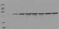
Detection of human Fos, phosphorylated on Ser374, by immunoblotting. Samples: Serum starved HepG2 cells incubated with EGF for 0 min (lane 1), 5 min (lane 2), 15 min (lane 3), 30 min (lane 4), 1 h (lane 5), 2 h (lane 6), 4 h (lane 7), and 8 h (lane 8). Primary antibody: PhosphoDetect™ Anti-Fos (pSer374) Mouse mAb (34E4) (Cat. No. ST1029) (0.5 µg/ml). Detection: chemiluminescence.
References
References
Chen, R-H., et al. 1993. Proc. Natl. Acad. Sci. USA90, 10952.
Product Information
Form
Lyophilized
Formulation
100 µg antibody lyophilized from 2X PBS, PEG, sucrose and 200 µl lyophilized control lysate from EGF-treated HEPG2 cells.
Does not detect the unphosphorylated form of c-fos. For immunoblotting use 20 µl control lysate per lane (minigel) for chemiluminescent detection. Antibody should be titrated for optimal results in individual systems.
Biological Information
Immunogen
a synthetic phosphopeptide corresponding to amino acids surrounding the Ser³⁷⁴ phosphorylation site of human c-Fos
Immunogen
Human
Clone
34E4
Host
Mouse
Isotype
IgG₁
Species Reactivity
Human
Antibody Type
Monoclonal Antibody
Physicochemical Information
Dimensions
Materials Information
Toxicological Information
Safety Information according to GHS
Safety Information
Product Usage Statements
Storage and Shipping Information
Ship Code
Shipped with Blue Ice or with Dry Ice
Toxicity
Irritant
Storage
-20°C
Avoid freeze/thaw
Avoid freeze/thaw
Do not freeze
Ok to freeze
Special Instructions
Reconstitute antibody with 1 ml water (15 minutes, room temperature). Following reconstitution, aliquot and freeze in liquid nitrogen. Reconstituted antibody can be stored at -80°C for up to 1 year. Aliquots may be stored at 4°C for up to 3 months and should be thawed at 37°C. Reconstitute control lysate with 200 µl H₂O. After complete solubilization of the proteins add 200 µl SDS-PAGE sample buffer and incubate at 90°C for 5 min. Following reconsitution aliquot and freeze (-20°C). Avoid freeze/thaw cycles.
Packaging Information
Transport Information
Supplemental Information
Specifications
Global Trade Item Number
Bestellnummer
GTIN
ST1029-1SET
04055977208689
Documentation
PhosphoDetect™ Anti-Fos (pSer³⁷⁴) Mouse mAb (34E4) SDB
PhosphoDetect™ Anti-Fos (pSer³⁷⁴) Mouse mAb (34E4) Analysenzertifikate
Titel
Chargennummer
ST1029
Literatur
Übersicht
Chen, R-H., et al. 1993. Proc. Natl. Acad. Sci. USA90, 10952.
Datenblatt
Note that this data sheet is not lot-specific and is representative of the current specifications for this product. Please consult the vial label and the certificate of analysis for information on specific lots. Also note that shipping conditions may differ from storage conditions.
Detection of human Fos, phosphorylated on Ser374, by immunoblotting. Samples: Serum starved HepG2 cells incubated with EGF for 0 min (lane 1), 5 min (lane 2), 15 min (lane 3), 30 min (lane 4), 1 h (lane 5), 2 h (lane 6), 4 h (lane 7), and 8 h (lane 8). Primary antibody: PhosphoDetect™ Anti-Fos (pSer374) Mouse mAb (34E4) (Cat. No. ST1029) (0.5 µg/ml). Detection: chemiluminescence.
Description
Mouse monoclonal antibody purified from serum-free cell culture supernatant by thiophilic adsorption and size exclusion chromatography. Supplied with a positive control lysate consisting of EGF-treated HepG2 cells. Recognizes the ~55 kDa c-fos protein phosphorylated at Ser374 by MAP kinase.
Background
The early gene product c-Fos is expressed following mitogenic stimulation and functions as a sensor for MAPK signal duration. When MAPK activation is transient, MAPK activity declines before accumulation of the c-Fos protein. When MAPK activation is sustained, c-Fos is phosphorylated by MAPK at Ser374. Phosphorylation stabilizes the Fos protein and primes c-Fos for additional phosphorylation at Thr325.
Host
Mouse
Immunogen species
Human
Immunogen
a synthetic phosphopeptide corresponding to amino acids surrounding the Ser³⁷⁴ phosphorylation site of human c-Fos
Clone
34E4
Isotype
IgG₁
Species
human
Positive control
HepG2 cells treated with EGF
Form
Lyophilized
Formulation
100 µg antibody lyophilized from 2X PBS, PEG, sucrose and 200 µl lyophilized control lysate from EGF-treated HEPG2 cells.
Preservative
≤0.1% sodium azide (antibody only)
Comments
Does not detect the unphosphorylated form of c-fos. For immunoblotting use 20 µl control lysate per lane (minigel) for chemiluminescent detection. Antibody should be titrated for optimal results in individual systems.
Storage
Avoid freeze/thaw
-20°C
Do Not Freeze
Ok to freeze
Special Instructions
Reconstitute antibody with 1 ml water (15 minutes, room temperature). Following reconstitution, aliquot and freeze in liquid nitrogen. Reconstituted antibody can be stored at -80°C for up to 1 year. Aliquots may be stored at 4°C for up to 3 months and should be thawed at 37°C. Reconstitute control lysate with 200 µl H₂O. After complete solubilization of the proteins add 200 µl SDS-PAGE sample buffer and incubate at 90°C for 5 min. Following reconsitution aliquot and freeze (-20°C). Avoid freeze/thaw cycles.
Toxicity
Irritant
References
Chen, R-H., et al. 1993. Proc. Natl. Acad. Sci. USA90, 10952.