Wenn Sie das Fenster schließen, wird Ihre Konfiguration nicht gespeichert, es sei denn, Sie haben Ihren Artikel in die Bestellung aufgenommen oder zu Ihren Favoriten hinzugefügt.
Klicken Sie auf OK, um das MILLIPLEX® MAP-Tool zu schließen oder auf Abbrechen, um zu Ihrer Auswahl zurückzukehren.
Wählen Sie konfigurierbare Panels & Premixed-Kits - ODER - Kits für die zelluläre Signaltransduktion & MAPmates™
Konfigurieren Sie Ihre MILLIPLEX® MAP-Kits und lassen sich den Preis anzeigen.
Konfigurierbare Panels & Premixed-Kits
Unser breites Angebot enthält Multiplex-Panels, für die Sie die Analyten auswählen können, die am besten für Ihre Anwendung geeignet sind. Unter einem separaten Register können Sie das Premixed-Cytokin-Format oder ein Singleplex-Kit wählen.
Kits für die zelluläre Signaltransduktion & MAPmates™
Wählen Sie gebrauchsfertige Kits zur Erforschung gesamter Signalwege oder Prozesse. Oder konfigurieren Sie Ihre eigenen Kits mit Singleplex MAPmates™.
Die folgenden MAPmates™ sollten nicht zusammen analysiert werden: -MAPmates™, die einen unterschiedlichen Assaypuffer erfordern. -Phosphospezifische und MAPmate™ Gesamtkombinationen wie Gesamt-GSK3β und Gesamt-GSK3β (Ser 9). -PanTyr und locusspezifische MAPmates™, z.B. Phospho-EGF-Rezeptor und Phospho-STAT1 (Tyr701). -Mehr als 1 Phospho-MAPmate™ für ein einziges Target (Akt, STAT3). -GAPDH und β-Tubulin können nicht mit Kits oder MAPmates™, die panTyr enthalten, analysiert werden.
.
Bestellnummer
Bestellinformationen
St./Pkg.
Liste
Dieser Artikel wurde zu Ihren Favoriten hinzugefügt.
Wählen Sie bitte Spezies, Panelart, Kit oder Probenart
Um Ihr MILLIPLEX® MAP-Kit zu konfigurieren, wählen Sie zunächst eine Spezies, eine Panelart und/oder ein Kit.
Custom Premix Selecting "Custom Premix" option means that all of the beads you have chosen will be premixed in manufacturing before the kit is sent to you.
Catalogue Number
Ordering Description
Qty/Pack
List
Dieser Artikel wurde zu Ihren Favoriten hinzugefügt.
Spezies
Panelart
Gewähltes Kit
Menge
Bestellnummer
Bestellinformationen
St./Pkg.
Listenpreis
96-Well Plate
Menge
Bestellnummer
Bestellinformationen
St./Pkg.
Listenpreis
Weitere Reagenzien hinzufügen (MAPmates erfordern die Verwendung eines Puffer- und Detektionskits)
Menge
Bestellnummer
Bestellinformationen
St./Pkg.
Listenpreis
48-602MAG
Buffer Detection Kit for Magnetic Beads
1 Kit
Platzsparende Option Kunden, die mehrere Kits kaufen, können ihre Multiplex-Assaykomponenten in Kunststoffbeuteln anstelle von Packungen erhalten, um eine kompaktere Lagerung zu ermöglichen.
Dieser Artikel wurde zu Ihren Favoriten hinzugefügt.
Das Produkt wurde in Ihre Bestellung aufgenommen
Sie können nun ein weiteres Kit konfigurieren, ein Premixed-Kit wählen, zur Kasse gehen oder das Bestell-Tool schließen.
17-10387
Sigma-AldrichLentiBrite™ GFP Control Lentiviral Biosensor
LentiBrite™ GFP Control Lentiviral Biosensor: SDB (Sicherheitsdatenblätter), Analysenzertifikate und Qualitätszertifikate, Dossiers, Broschüren und andere verfügbare Dokumente.
Glutathione, Oxidized, Free Acid - CAS 27025-41-8 - Calbiochem
Übersicht
Replacement Information
Key Spec Table
Key Applications
Detection Methods
TFX, IF, ICC
Fluorescent
Description
Catalogue Number
17-10387
Trade Name
LentiBrite
Chemicon
Description
LentiBrite™ GFP Control Lentiviral Biosensor
Overview
Read our application note in Nature Methods! http://www.nature.com/app_notes/nmeth/2012/121007/pdf/an8620.pdf (Click Here!)
Learn more about the advantages of our LentiBrite Lentiviral Biosensors! Click Here
Biosensors can be used to detect the presence/absence of a particular protein as well as the subcellular location of that protein within the live state of a cell. Fluorescent tags are often desired as a means to visualize the protein of interest within a cell by either fluorescent microscopy or time-lapse video capture. Visualizing live cells without disruption allows researchers to observe cellular conditions in real time.
Lentiviral vector systems are a popular research tool used to introduce gene products into cells. Lentiviral transfection has advantages over non-viral methods such as chemical-based transfection including higher-efficiency transfection of dividing and non-dividing cells, long-term stable expression of the transgene, and low immunogenicity.
EMD Millipore is introducing LentiBrite™ Lentiviral Biosensors, a new suite of pre-packaged lentiviral particles encoding important and foundational proteins of autophagy, apoptosis, and cell structure for visualization under different cell/disease states in live cell and in vitro analysis.
Pre-packaged, fluorescently-tagged with GFP & RFP
Higher efficiency transfection as compared to traditional chemical-based and other non-viral-based transfection methods
Ability to transfect dividing, non-dividing, and difficult-to-transfect cell types, such as primary cells or stem cells
Non-disruptive towards cellular function
EMD Millipore’s LentiBrite™ GFP Control lentiviral particles provide bright fluorescence to enable live-cell analysis in difficult-to-transfect cell types.
Background Information
The use of genetically encoded fluorescent proteins has revolutionized cell biology by enabling real-time imaging of cells and subcellular structures. A diverse palette of fluorescent proteins is available, but these proteins vary in their suitability for live-cell imaging. EMD Millipore’s LentiBriteTM GFP Control Lentiviral Biosensor employs TagGFP2, the improved variant of the Aequorea macrodactyla GFP-like protein. TagGFP2 exhibits bright green fluorescence comparable to that of EGFP, with excitation/emission maxima at 483 and 506 nm, respectively. TagGFP2 is specially optimized for expression at 37°C and is therefore well-suited for use in mammalian cells. EMD Millipore’s LentiBrite™ GFP Control lentiviral particles provide bright fluorescence to enable live-cell analysis in difficult-to-transfect cell types.
References
Product Information
Components
TagGFP2 Lentivirus: One vial containing 25 µL of lentiviral particles at a minimum of 3 x 10E8 infectious units (IFU) per mL. For lot-specific titer information, please see lot specific “Viral Titer” in the product specifications of the datasheet.
Promoter EF-1 (Elongation Factor-1)
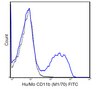
Multiplicty of Infection (MOI) MOI = Ratio of # of infectious lentiviral particles (IFU) to # of cells being infected. Typical MOI values for high transduction efficiency and signal intensity are in the range of 20-40. For this target, some cell types may require lower MOIs (e.g., HT-1080, HeLa, U2OS, human mesenchymal stem cells (HuMSC)), while others may require higher MOIs (e.g., human umbilical vein endothelial cells (HUVEC)). NOTE: MOI should be titrated and optimized by the end user for each cell type and lentiviral target to achieve desired transduction efficiency and signal intensity.
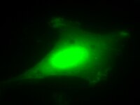
Fluorescence Microscopy Imaging: (See Figure 1 in datasheet) HeLa cells were plated in a chamber slide and transduced with lentiviral particles at an MOI of 40 for 24 hours. After media replacement and 48 hours further incubation, cells were fixed with formaldehyde and mounted. Images were obtained by oil immersion wide-field fluorescence microscopy. The GFP displays a uniform green fluorescent distribution in the cells.
Immunocytochemistry Comparison: (See Figure 2 in datasheet) Similar to Figure 1 (see datasheet), U2OS cells were plated in a chamber slide and transduced with lentiviral particles at an MOI of 40 for 24 hours. After 24 hours, media was replaced and cells were then further incubated for 48 hours.
Hard-to-transfect Cell Types: (See Figure 3 in datasheet) Primary cell types HUVEC or HuMSC were plated in chamber slides and transduced with lentiviral particles at an MOI of 40 for 24 hours.
Additional cell type: (See Figure 4 in datasheet) Primary Rat Cortical Neuron cells were treated as in Figure 1 (see datasheet).
For optimal fluorescent visualization, it is recommended to analyze the target expression level within 24-48 hrs after transfection/infection for optimal live cell analysis, as fluorescent intensity may dim over time, especially in difficult-to-transfect cell lines. Infected cells may be frozen down after successful transfection/infection and thawed in culture to retain positive fluorescent expression beyond 24-48 hrs. Length and intensity of fluorescent expression varies between cell lines. Higher MOIs may be required for difficult-to-transfect cell lines.
Biological Information
Purification Method
PEG precipitation
Physicochemical Information
Dimensions
Materials Information
Toxicological Information
Safety Information according to GHS
Safety Information
Product Usage Statements
Quality Assurance
Evaluated by transduction of HT-1080 cells and fluorescent imaging performed for assessment of transduction efficiency.
Usage Statement
This product contains genetically modified organisms (GMO). Within the EU GMOs are regulated by Directives 2001/18/EC and 2009/41/EC of the European Parliament and of the Council and their national implementation in the member States respectively. This legislation obliges Merck to request certain information about you and the establishment where the GMOs are being handled. Click here for Enduser Declaration (EUD) Form.
Unless otherwise stated in our catalog or other company documentation accompanying the product(s), our products are intended for research use only and are not to be used for any other purpose, which includes but is not limited to, unauthorized commercial uses, in vitro diagnostic uses, ex vivo or in vivo therapeutic uses or any type of consumption or application to humans or animals.
Storage and Shipping Information
Storage Conditions
Storage and Handling Lentivirus is stable for at least 4 months from date of receipt when stored at -80°C. After first thaw, place immediately on ice and freeze in working aliquots at -80°C. Frozen aliquots may be stored for at least 2 months. Further freeze/thaws may result in decreased virus titer and transduction efficiency.
IMPORTANT SAFETY NOTE Replication-defective lentiviral vectors, such as the 3rd Generation vector provided in this product, are not known to cause any diseases in humans or animals. However, lentiviruses can integrate into the host cell genome and thus pose some risk of insertional mutagenesis. Material is a Risk Group 2 and should be handled under BSL2 controls. A detailed discussion of biosafety of lentiviral vectors is provided in Pauwels, K. et al. (2009). State-of-the-art lentiviral vectors for research use: Risk assessment and biosafety recommendations. Curr. Gene Ther. 9: 459-474.