Wenn Sie das Fenster schließen, wird Ihre Konfiguration nicht gespeichert, es sei denn, Sie haben Ihren Artikel in die Bestellung aufgenommen oder zu Ihren Favoriten hinzugefügt.
Klicken Sie auf OK, um das MILLIPLEX® MAP-Tool zu schließen oder auf Abbrechen, um zu Ihrer Auswahl zurückzukehren.
Wählen Sie konfigurierbare Panels & Premixed-Kits - ODER - Kits für die zelluläre Signaltransduktion & MAPmates™
Konfigurieren Sie Ihre MILLIPLEX® MAP-Kits und lassen sich den Preis anzeigen.
Konfigurierbare Panels & Premixed-Kits
Unser breites Angebot enthält Multiplex-Panels, für die Sie die Analyten auswählen können, die am besten für Ihre Anwendung geeignet sind. Unter einem separaten Register können Sie das Premixed-Cytokin-Format oder ein Singleplex-Kit wählen.
Kits für die zelluläre Signaltransduktion & MAPmates™
Wählen Sie gebrauchsfertige Kits zur Erforschung gesamter Signalwege oder Prozesse. Oder konfigurieren Sie Ihre eigenen Kits mit Singleplex MAPmates™.
Die folgenden MAPmates™ sollten nicht zusammen analysiert werden: -MAPmates™, die einen unterschiedlichen Assaypuffer erfordern. -Phosphospezifische und MAPmate™ Gesamtkombinationen wie Gesamt-GSK3β und Gesamt-GSK3β (Ser 9). -PanTyr und locusspezifische MAPmates™, z.B. Phospho-EGF-Rezeptor und Phospho-STAT1 (Tyr701). -Mehr als 1 Phospho-MAPmate™ für ein einziges Target (Akt, STAT3). -GAPDH und β-Tubulin können nicht mit Kits oder MAPmates™, die panTyr enthalten, analysiert werden.
.
Bestellnummer
Bestellinformationen
St./Pkg.
Liste
Dieser Artikel wurde zu Ihren Favoriten hinzugefügt.
Wählen Sie bitte Spezies, Panelart, Kit oder Probenart
Um Ihr MILLIPLEX® MAP-Kit zu konfigurieren, wählen Sie zunächst eine Spezies, eine Panelart und/oder ein Kit.
Custom Premix Selecting "Custom Premix" option means that all of the beads you have chosen will be premixed in manufacturing before the kit is sent to you.
Catalogue Number
Ordering Description
Qty/Pack
List
Dieser Artikel wurde zu Ihren Favoriten hinzugefügt.
Spezies
Panelart
Gewähltes Kit
Menge
Bestellnummer
Bestellinformationen
St./Pkg.
Listenpreis
96-Well Plate
Menge
Bestellnummer
Bestellinformationen
St./Pkg.
Listenpreis
Weitere Reagenzien hinzufügen (MAPmates erfordern die Verwendung eines Puffer- und Detektionskits)
Menge
Bestellnummer
Bestellinformationen
St./Pkg.
Listenpreis
48-602MAG
Buffer Detection Kit for Magnetic Beads
1 Kit
Platzsparende Option Kunden, die mehrere Kits kaufen, können ihre Multiplex-Assaykomponenten in Kunststoffbeuteln anstelle von Packungen erhalten, um eine kompaktere Lagerung zu ermöglichen.
Dieser Artikel wurde zu Ihren Favoriten hinzugefügt.
Das Produkt wurde in Ihre Bestellung aufgenommen
Sie können nun ein weiteres Kit konfigurieren, ein Premixed-Kit wählen, zur Kasse gehen oder das Bestell-Tool schließen.
SCC211
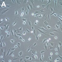
Sigma-AldrichIngWAT Mouse Preadipocyte Cell Line
The IngWAT mouse immortalized preadipocyte cell line is derived from subcutaneous white adipose tissue (WAT) and has been shown to support the induction of beige adipocytes that express UCP1.
More>>The IngWAT mouse immortalized preadipocyte cell line is derived from subcutaneous white adipose tissue (WAT) and has been shown to support the induction of beige adipocytes that express UCP1. Less<<
Empfohlene Produkte
Übersicht
Replacement Information
Description
Catalogue Number
SCC211
Description
IngWAT Mouse Preadipocyte Cell Line
Overview
Source: Stromal vascular fractions were isolated from the inguinal white adipose tissue of a wild-type mouse and immortalized by infection with Simian virus 40 (SV40) large T antigen (2).
Background Information
The growing worldwide obesity epidemic has stimulated research into novel strategies to control and treat obesity and associated metabolic disorders. Great interest has been generated in brown fat, a specialized adipose tissue having increased energy expenditure, because manipulation of this tissue has potential to protect against diet-induced obesity by altering the balance of energy intake and consumption (1). In common with brown fat, beige adipocytes possess thermogenic mechanisms, but have the unique property of inducible differentiation. Beige adipocyte differentiation occurs within white adipose tissue in response to environmental cues and is characterized by cristae-dense mitochondrial biogenesis and increased expression of uncoupling protein 1 (UCP1) (2).
The IngWAT mouse immortalized preadipocyte cell line is derived from subcutaneous white adipose tissue (WAT) isolated from a wild-type mouse (2). IngWAT cells have been shown to support the induction of beige adipocytes and express UCP1.2 IngWAT mouse immortalized preadipocyte cells are a versatile model for a broad spectrum of studies of beige adipocyte induction, maintenance and physiology.
References: 1. Cui XB, Chen SY (2016) White adipose tissue browning and obesity. 31(1): 1-2. 2. Lu X et al. (2018) Mitophagy controls beige adipocyte maintenance through a Parkin-dependent and UCP1-independent mechanism. Sci Signal 11(527): eaap8526.
References
Product Information
Applications
Application
The IngWAT mouse immortalized preadipocyte cell line is derived from subcutaneous white adipose tissue (WAT) and has been shown to support the induction of beige adipocytes that express UCP1.
Key Applications
Cell Culture
Application Notes
This product is intended for sale and sold solely to academic institutions for internal academic research use per the terms of the “Academic Use Agreement” as detailed in the product documentation. For information regarding any other use, please contact licensing@emdmillipore.com.
Biological Information
Cell Line Type
Adipocytes
Physicochemical Information
Dimensions
Materials Information
Toxicological Information
Safety Information according to GHS
Safety Information
Product Usage Statements
Quality Assurance
• Each vial contains ≥ 1X10⁶ viable cells. • Cells are tested negative for infectious diseases by a Mouse Essential CLEAR panel by Charles River Animal Diagnostic Services. • Cells are verified to be of mouse origin and negative for inter-species contamination from rat, chinese hamster, Golden Syrian hamster, human and non-human primate (NHP) as assessed by a Contamination CLEAR panel by Charles River Animal Diagnostic Services. • Cells are negative for mycoplasma contamination.
Usage Statement
This product contains genetically modified organisms (GMO). Within the EU GMOs are regulated by Directives 2001/18/EC and 2009/41/EC of the European Parliament and of the Council and their national implementation in the member States respectively. This legislation obliges Merck to request certain information about you and the establishment where the GMOs are being handled. Click here for Enduser Declaration (EUD) Form.
Unless otherwise stated in our catalog or other company documentation accompanying the product(s), our products are intended for research use only and are not to be used for any other purpose, which includes but is not limited to, unauthorized commercial uses, in vitro diagnostic uses, ex vivo or in vivo therapeutic uses or any type of consumption or application to humans or animals.
Storage and Shipping Information
Storage Conditions
Store in liquid nitrogen. The cells can be cultured for at least 10 passages after initial thawing without significantly affecting the cell marker expression and functionality.