Wenn Sie das Fenster schließen, wird Ihre Konfiguration nicht gespeichert, es sei denn, Sie haben Ihren Artikel in die Bestellung aufgenommen oder zu Ihren Favoriten hinzugefügt.
Klicken Sie auf OK, um das MILLIPLEX® MAP-Tool zu schließen oder auf Abbrechen, um zu Ihrer Auswahl zurückzukehren.
Wählen Sie konfigurierbare Panels & Premixed-Kits - ODER - Kits für die zelluläre Signaltransduktion & MAPmates™
Konfigurieren Sie Ihre MILLIPLEX® MAP-Kits und lassen sich den Preis anzeigen.
Konfigurierbare Panels & Premixed-Kits
Unser breites Angebot enthält Multiplex-Panels, für die Sie die Analyten auswählen können, die am besten für Ihre Anwendung geeignet sind. Unter einem separaten Register können Sie das Premixed-Cytokin-Format oder ein Singleplex-Kit wählen.
Kits für die zelluläre Signaltransduktion & MAPmates™
Wählen Sie gebrauchsfertige Kits zur Erforschung gesamter Signalwege oder Prozesse. Oder konfigurieren Sie Ihre eigenen Kits mit Singleplex MAPmates™.
Die folgenden MAPmates™ sollten nicht zusammen analysiert werden: -MAPmates™, die einen unterschiedlichen Assaypuffer erfordern. -Phosphospezifische und MAPmate™ Gesamtkombinationen wie Gesamt-GSK3β und Gesamt-GSK3β (Ser 9). -PanTyr und locusspezifische MAPmates™, z.B. Phospho-EGF-Rezeptor und Phospho-STAT1 (Tyr701). -Mehr als 1 Phospho-MAPmate™ für ein einziges Target (Akt, STAT3). -GAPDH und β-Tubulin können nicht mit Kits oder MAPmates™, die panTyr enthalten, analysiert werden.
.
Bestellnummer
Bestellinformationen
St./Pkg.
Liste
Dieser Artikel wurde zu Ihren Favoriten hinzugefügt.
Wählen Sie bitte Spezies, Panelart, Kit oder Probenart
Um Ihr MILLIPLEX® MAP-Kit zu konfigurieren, wählen Sie zunächst eine Spezies, eine Panelart und/oder ein Kit.
Custom Premix Selecting "Custom Premix" option means that all of the beads you have chosen will be premixed in manufacturing before the kit is sent to you.
Catalogue Number
Ordering Description
Qty/Pack
List
Dieser Artikel wurde zu Ihren Favoriten hinzugefügt.
Spezies
Panelart
Gewähltes Kit
Menge
Bestellnummer
Bestellinformationen
St./Pkg.
Listenpreis
96-Well Plate
Menge
Bestellnummer
Bestellinformationen
St./Pkg.
Listenpreis
Weitere Reagenzien hinzufügen (MAPmates erfordern die Verwendung eines Puffer- und Detektionskits)
Menge
Bestellnummer
Bestellinformationen
St./Pkg.
Listenpreis
48-602MAG
Buffer Detection Kit for Magnetic Beads
1 Kit
Platzsparende Option Kunden, die mehrere Kits kaufen, können ihre Multiplex-Assaykomponenten in Kunststoffbeuteln anstelle von Packungen erhalten, um eine kompaktere Lagerung zu ermöglichen.
Dieser Artikel wurde zu Ihren Favoriten hinzugefügt.
Das Produkt wurde in Ihre Bestellung aufgenommen
Sie können nun ein weiteres Kit konfigurieren, ein Premixed-Kit wählen, zur Kasse gehen oder das Bestell-Tool schließen.
This phospho-PKD2 antibody is validated for use in WB for the detection of the phospho-PKD2 protein.
More>>This phospho-PKD2 antibody is validated for use in WB for the detection of the phospho-PKD2 protein. Less<<
Anti-phospho-PKD2 Antibody (Ser197/Ser200): SDB (Sicherheitsdatenblätter), Analysenzertifikate und Qualitätszertifikate, Dossiers, Broschüren und andere verfügbare Dokumente.
Autosomal dominant polycystic kidney disease type II protein
Polycystic kidney disease 2 protein
Polycystwin
R48321
Background Information
As members of the calcium-/calmodulin-depedent protein kinase superfamily, there are three known Protein Kinase D (PKD) isoforms, PKD1, PKD2, and PKD3. These serine/threonine kinases are involved in protein-protein and protein-carbohydrate adhesion and are also thought to be ion channel regulators. They are prominent downstream targets of protein kinase C and phospholipase D in various biological systems including mouse. PKDs are activated by various stimuli, including phorbol esters, G-protein-coupled receptors, reactive oxygen species, and diacylglycerol. PKDs regulate protein transport in the trans-Golgi network and are thought to play a role in muscle differentiation, cell motility, migration, and invasion. These isoforms are known to interact with each other. PKD2 is shown to be activated by phorbol esters, both in vivo and in vitro, and also by gastrin via the cholescystokinin/CCK(B) receptor in stably-transfected AGS-B cells. Furthermore, PKD2 is found to be active during the early phase of differentation of C2C12 cells into skeletal muscle cells. It is also reported to enhance TCR-induced NFAT activity in Jurkat T cells.
References
Product Information
Format
Affinity Purified
Control
MEF cell lysates untreated and PBDu-treated
Presentation
Purified rabbit polyclonal in buffer containing 0.1 M Tris-Glycine (pH 7.4), 150 mM NaCl with 0.05% sodium azide.
Applications
Application
This phospho-PKD2 antibody is validated for use in WB for the detection of the phospho-PKD2 protein.
Key Applications
Western Blotting
Application Notes
Evaluated by Western Blot in MEF cell lysates untreated and treated with PDBu, Rapamycin, and GO6983.
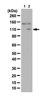
Western Blot Analysis: A 1:2,000 dilution of this antibody detected PKD2 in 10 µg of MEF cell lysates untreated and treated with PDBu, Rapamycin, and GO6983.
Biological Information
Immunogen
KLH-conjugated linear peptide corresponding to human PKD2 phosphorylated at Ser197/Ser200.
Epitope
Phosphorylated Ser197/Ser200
Concentration
Please refer to the Certificate of Analysis for the lot-specific concentration.
Host
Rabbit
Specificity
This antibody recognizes PKD2 phosphorylated at Ser197/Ser200.
Species Reactivity
Mouse
Human
Rat
Species Reactivity Note
Demonstrated to react with Mouse. Predicted to react with Human and Rat based on 100% sequence homology.
This gene encodes a member of the polycystin protein family. The encoded protein is a multi-pass membrane protein that functions as a calcium permeable cation channel, and is involved in calcium transport and calcium signaling in renal epithelial cells. This protein interacts with polycystin 1, and they may be partners in a common signaling cascade involved in tubular morphogenesis. Mutations in this gene are associated with autosomal dominant polycystic kidney disease type 2.
FUNCTION: Functions as a calcium permeable cation channel. PKD1 and PKD2 may function through a common signaling pathway that is necessary for normal tubulogenesis.
SUBUNIT STRUCTURE: Forms homooligomers. Interacts with PKD1. PKD1 requires the presence of PKD2 for stable expression. Interacts with CD2AP. Interacts with HAX1.
SUBCELLULAR LOCATION: Multi-pass membrane protein Potential. Endoplasmic reticulum.
Molecular Weight
~110 kDa observed. An uncharacterized band appears at ~150 kDa in some lysates.
Physicochemical Information
Dimensions
Materials Information
Toxicological Information
Safety Information according to GHS
Safety Information
Product Usage Statements
Quality Assurance
Evaluated by Western Blot in MEF cell lysates untreated and PBDu-treated.
Western Blot Analysis: A 1:2,000 dilution of this antibody detected PKD2 in 10 µg of MEF cell lysates untreated and PBDu-treated.
Usage Statement
Unless otherwise stated in our catalog or other company documentation accompanying the product(s), our products are intended for research use only and are not to be used for any other purpose, which includes but is not limited to, unauthorized commercial uses, in vitro diagnostic uses, ex vivo or in vivo therapeutic uses or any type of consumption or application to humans or animals.