Wenn Sie das Fenster schließen, wird Ihre Konfiguration nicht gespeichert, es sei denn, Sie haben Ihren Artikel in die Bestellung aufgenommen oder zu Ihren Favoriten hinzugefügt.
Klicken Sie auf OK, um das MILLIPLEX® MAP-Tool zu schließen oder auf Abbrechen, um zu Ihrer Auswahl zurückzukehren.
Wählen Sie konfigurierbare Panels & Premixed-Kits - ODER - Kits für die zelluläre Signaltransduktion & MAPmates™
Konfigurieren Sie Ihre MILLIPLEX® MAP-Kits und lassen sich den Preis anzeigen.
Konfigurierbare Panels & Premixed-Kits
Unser breites Angebot enthält Multiplex-Panels, für die Sie die Analyten auswählen können, die am besten für Ihre Anwendung geeignet sind. Unter einem separaten Register können Sie das Premixed-Cytokin-Format oder ein Singleplex-Kit wählen.
Kits für die zelluläre Signaltransduktion & MAPmates™
Wählen Sie gebrauchsfertige Kits zur Erforschung gesamter Signalwege oder Prozesse. Oder konfigurieren Sie Ihre eigenen Kits mit Singleplex MAPmates™.
Die folgenden MAPmates™ sollten nicht zusammen analysiert werden: -MAPmates™, die einen unterschiedlichen Assaypuffer erfordern. -Phosphospezifische und MAPmate™ Gesamtkombinationen wie Gesamt-GSK3β und Gesamt-GSK3β (Ser 9). -PanTyr und locusspezifische MAPmates™, z.B. Phospho-EGF-Rezeptor und Phospho-STAT1 (Tyr701). -Mehr als 1 Phospho-MAPmate™ für ein einziges Target (Akt, STAT3). -GAPDH und β-Tubulin können nicht mit Kits oder MAPmates™, die panTyr enthalten, analysiert werden.
.
Bestellnummer
Bestellinformationen
St./Pkg.
Liste
Dieser Artikel wurde zu Ihren Favoriten hinzugefügt.
Wählen Sie bitte Spezies, Panelart, Kit oder Probenart
Um Ihr MILLIPLEX® MAP-Kit zu konfigurieren, wählen Sie zunächst eine Spezies, eine Panelart und/oder ein Kit.
Custom Premix Selecting "Custom Premix" option means that all of the beads you have chosen will be premixed in manufacturing before the kit is sent to you.
Catalogue Number
Ordering Description
Qty/Pack
List
Dieser Artikel wurde zu Ihren Favoriten hinzugefügt.
Spezies
Panelart
Gewähltes Kit
Menge
Bestellnummer
Bestellinformationen
St./Pkg.
Listenpreis
96-Well Plate
Menge
Bestellnummer
Bestellinformationen
St./Pkg.
Listenpreis
Weitere Reagenzien hinzufügen (MAPmates erfordern die Verwendung eines Puffer- und Detektionskits)
Menge
Bestellnummer
Bestellinformationen
St./Pkg.
Listenpreis
48-602MAG
Buffer Detection Kit for Magnetic Beads
1 Kit
Platzsparende Option Kunden, die mehrere Kits kaufen, können ihre Multiplex-Assaykomponenten in Kunststoffbeuteln anstelle von Packungen erhalten, um eine kompaktere Lagerung zu ermöglichen.
Dieser Artikel wurde zu Ihren Favoriten hinzugefügt.
Das Produkt wurde in Ihre Bestellung aufgenommen
Sie können nun ein weiteres Kit konfigurieren, ein Premixed-Kit wählen, zur Kasse gehen oder das Bestell-Tool schließen.
ABS1646
Sigma-AldrichAnti-VPS4A (UT289)
Detect Vacuolar protein sorting-associated protein 4A using this rabbit polyclonal Anti-VPS4A (UT289), Cat. No. ABS1646, validated for use in Immunocytochemistry, Immunoprecipitation, and Western Blotting.
More>>Detect Vacuolar protein sorting-associated protein 4A using this rabbit polyclonal Anti-VPS4A (UT289), Cat. No. ABS1646, validated for use in Immunocytochemistry, Immunoprecipitation, and Western Blotting. Less<<
Anti-VPS4A (UT289): SDB (Sicherheitsdatenblätter), Analysenzertifikate und Qualitätszertifikate, Dossiers, Broschüren und andere verfügbare Dokumente.
Vacuolar protein sorting-associated protein 4A (EC 3.6.4.6; UniProt Q9UN37; also known as hVPS4, Protein SKD2, SKD1-homolog, Vacuolar protein sorting factor 4A, Vacuolar sorting protein 4, VPS4A) is encoded by the VPS4A (also known as SKD1A, VPS4, VPS4-1) gene (Gene ID 27183) in human. VPS4A is a member of the ATPases associated with various cellular activities (AAA) family proteins. The ESCRT (endosomal sorting complexes required for transport) machinery consists of five complexes (SCRT-0, ESCRT-I, ESCRT-II, ESCRT-III, and Vps4) and mediates multiple cellular membrane remodeling events, ranging from multivesicular bodies and exosomes formation to the final membrane abscission stage of cytokinesis, all of which converge in the scission of the narrow membrane neck that connects bud to parent membrane or daughter cell to daughter cell. Scission depends on the polymerization of soluble ESCRT-III monomers or dimers into tightly membrane-bound filaments. VPS4A is essential for the functions of the ESCRTs in cells by mediating the disassembly of ESCRT-III filaments to ensure a constant replenish of the pool of soluble ESCRT-III monomers. The ESCRTs are also exploited by HIV-1 and many other enveloped viruses for their escape from the host cells. In addition, VPS4A is reported to facilitate the secretion of oncogenic miRNAs in exosomes and downregulation of VPS4A in hepatocellular carcinoma (HCC) is associated with tumor progression and metastasis.
References
Product Information
Format
Serum
Presentation
Rabbit polyclonal antibody serum with 0.05% sodium azide.
Detect Vacuolar protein sorting-associated protein 4A using this rabbit polyclonal Anti-VPS4A (UT289), Cat. No. ABS1646, validated for use in Immunocytochemistry, Immunoprecipitation, and Western Blotting.
Key Applications
Immunocytochemistry
Western Blotting
Immunoprecipitation
Application Notes
Immunocytochemistry Analysis: A representative lot detected VPS4A immunoreactivity concentrated at the spindle pole during metaphase and at the midbody during cytokinesis in 3% paraformaldehyde-fixed HeLa cells. No staining was seen in VPS4A shRNA-transfected HeLa cells (Morita, E., et al. (2010). Proc. Natl. Acad. Sci. U.S.A. 107(29):12889-12894).
Immunoprecipitation Analysis: A representative lot immunoprecipited EGFP-VPS4A fusion together with CHMP5-Myc and Myc-LIP5 exogenously expressed in HEK293T cells. CHMP5-Myc with L4D mutation and Myc-LIP5 with M64A, W147D, or Y278A mutation failed to co-immunoprecipitate with VPS4A (Skalicky, J.J., et al. (2012). J. Biol. Chem. 287(52):43910-43926).
Western Blotting Analysis: A representative lot detected both endogenous VPS4A and exogenously expressed EGFP-VPS4A fusion in HEK293T cell lysates as well as in Anti-Myc immunoprecipites from CHMP5-Myc or Myc-LIP5 expressing cells (Skalicky, J.J., et al. (2012). J. Biol. Chem. 287(52):43910-43926).
Western Blotting Analysis: A representative lot detected the VPS4A target band in lysate from untransfected, but not VPS4A shRNA-transfected, HeLa cells (Morita, E., et al. (2010). Proc. Natl. Acad. Sci. U.S.A. 107(29):12889-12894).
Biological Information
Immunogen
Recombinant human VPS4A lacking N-terminal MIT domain (Morita, E., et al. (2010). Proc. Natl. Acad. Sci. U.S.A. 107(29):12889-12894).
Concentration
Please refer to lot specific datasheet.
Host
Rabbit
Specificity
VPS4A immunostaining and target band detection by UT289 was observed only in untransfected, but not VPS4A shRNA-transfected HeLa cells (Morita, E., et al. (2010). Proc. Natl. Acad. Sci. U.S.A. 107(29):12889-12894). Due to a typographical error, UT289 was listed as UT829 in Table S1 of Skalicky, J.J., et al. (2012). J. Biol. Chem. 287(52):43910-43926.
~52 kDa observed. 48.90/48.91 kDa (human/mouse) calculated. Uncharacterized bands may be observed in some lysate(s).
Physicochemical Information
Dimensions
Materials Information
Toxicological Information
Safety Information according to GHS
Safety Information
Product Usage Statements
Quality Assurance
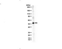
Evaluated by Western Blotting in MCF-7 cell lysate.
Western Blotting Analysis: A 1:1,000 dilution of this antiserum detected VPS4A in 10 µg of MCF-7 cell lysate.
Usage Statement
Unless otherwise stated in our catalog or other company documentation accompanying the product(s), our products are intended for research use only and are not to be used for any other purpose, which includes but is not limited to, unauthorized commercial uses, in vitro diagnostic uses, ex vivo or in vivo therapeutic uses or any type of consumption or application to humans or animals.
Storage and Shipping Information
Storage Conditions
Stable for 1 year at -20°C from date of receipt. Handling Recommendations: Upon receipt and prior to removing the cap, centrifuge the vial and gently mix the solution. Aliquot into microcentrifuge tubes and store at -20°C. Avoid repeated freeze/thaw cycles, which may damage IgG and affect product performance.