Wenn Sie das Fenster schließen, wird Ihre Konfiguration nicht gespeichert, es sei denn, Sie haben Ihren Artikel in die Bestellung aufgenommen oder zu Ihren Favoriten hinzugefügt.
Klicken Sie auf OK, um das MILLIPLEX® MAP-Tool zu schließen oder auf Abbrechen, um zu Ihrer Auswahl zurückzukehren.
Wählen Sie konfigurierbare Panels & Premixed-Kits - ODER - Kits für die zelluläre Signaltransduktion & MAPmates™
Konfigurieren Sie Ihre MILLIPLEX® MAP-Kits und lassen sich den Preis anzeigen.
Konfigurierbare Panels & Premixed-Kits
Unser breites Angebot enthält Multiplex-Panels, für die Sie die Analyten auswählen können, die am besten für Ihre Anwendung geeignet sind. Unter einem separaten Register können Sie das Premixed-Cytokin-Format oder ein Singleplex-Kit wählen.
Kits für die zelluläre Signaltransduktion & MAPmates™
Wählen Sie gebrauchsfertige Kits zur Erforschung gesamter Signalwege oder Prozesse. Oder konfigurieren Sie Ihre eigenen Kits mit Singleplex MAPmates™.
Die folgenden MAPmates™ sollten nicht zusammen analysiert werden: -MAPmates™, die einen unterschiedlichen Assaypuffer erfordern. -Phosphospezifische und MAPmate™ Gesamtkombinationen wie Gesamt-GSK3β und Gesamt-GSK3β (Ser 9). -PanTyr und locusspezifische MAPmates™, z.B. Phospho-EGF-Rezeptor und Phospho-STAT1 (Tyr701). -Mehr als 1 Phospho-MAPmate™ für ein einziges Target (Akt, STAT3). -GAPDH und β-Tubulin können nicht mit Kits oder MAPmates™, die panTyr enthalten, analysiert werden.
.
Bestellnummer
Bestellinformationen
St./Pkg.
Liste
Dieser Artikel wurde zu Ihren Favoriten hinzugefügt.
Wählen Sie bitte Spezies, Panelart, Kit oder Probenart
Um Ihr MILLIPLEX® MAP-Kit zu konfigurieren, wählen Sie zunächst eine Spezies, eine Panelart und/oder ein Kit.
Custom Premix Selecting "Custom Premix" option means that all of the beads you have chosen will be premixed in manufacturing before the kit is sent to you.
Catalogue Number
Ordering Description
Qty/Pack
List
Dieser Artikel wurde zu Ihren Favoriten hinzugefügt.
Spezies
Panelart
Gewähltes Kit
Menge
Bestellnummer
Bestellinformationen
St./Pkg.
Listenpreis
96-Well Plate
Menge
Bestellnummer
Bestellinformationen
St./Pkg.
Listenpreis
Weitere Reagenzien hinzufügen (MAPmates erfordern die Verwendung eines Puffer- und Detektionskits)
Menge
Bestellnummer
Bestellinformationen
St./Pkg.
Listenpreis
48-602MAG
Buffer Detection Kit for Magnetic Beads
1 Kit
Platzsparende Option Kunden, die mehrere Kits kaufen, können ihre Multiplex-Assaykomponenten in Kunststoffbeuteln anstelle von Packungen erhalten, um eine kompaktere Lagerung zu ermöglichen.
Dieser Artikel wurde zu Ihren Favoriten hinzugefügt.
Das Produkt wurde in Ihre Bestellung aufgenommen
Sie können nun ein weiteres Kit konfigurieren, ein Premixed-Kit wählen, zur Kasse gehen oder das Bestell-Tool schließen.
ABN1739-25UG
Sigma-AldrichAnti-Utrophin A
Anti-Utrophin A Antibody, Cat. No. ABN1739, is a highly specific rabbit polyclonal antibody that targets Utrophin and has been tested in Immunofluorescence, Immunohistochemistry (Paraffin), and Western Blotting.
More>>Anti-Utrophin A Antibody, Cat. No. ABN1739, is a highly specific rabbit polyclonal antibody that targets Utrophin and has been tested in Immunofluorescence, Immunohistochemistry (Paraffin), and Western Blotting. Less<<
Anti-Utrophin A: SDB (Sicherheitsdatenblätter), Analysenzertifikate und Qualitätszertifikate, Dossiers, Broschüren und andere verfügbare Dokumente.
EDTA, Tetrasodium Tetrahydrate Salt - CAS 13235-36-4 - Calbiochem
Übersicht
Replacement Information
Key Spec Table
Species Reactivity
Key Applications
Host
Format
Antibody Type
H, M
IF, IH(P), WB
Rb
Purified
Polyclonal Antibody
Description
Catalogue Number
ABN1739-25UG
Description
Anti-Utrophin A
Alternate Names
UTRN A
Background Information
Utrophin A (UniProt: E9Q6R7) is encoded by the Utrn gene in murine species. Utrophin is a large cytoskeletal protein, which is autosomally-encoded homologue of dystrophin, the protein product of the Duchenne muscular dystrophy (DMD) gene. It displays a sequence homology with dystrophin and possesses many of the protein-binding properties ascribed to dystrophin. In normal skeletal muscle, utrophin is located at the neuromuscular junction and dystrophin predominates at the sarcolemma. During development and in certain myopathies utrophin is also reported to be present at the sarcolemma and this redistribution is associated with higher levels of utrophin. Utrophin is found to co-localize with the acetylcholine receptors at the neuromuscular junctions and may participate in stabilizing the synaptic cytoskeleton. The utrophin mRNA contains two full-length species (named A- and B-utrophin), which are transcribed from different promoters. Hence, utrophin is a composite of A- and B-utrophin and only the A-utrophin is up-regulated in dystrophin-deficient striated muscle. This up-regulation is reported to occur post-transcriptionally. Utrophin A and B differ in their N-termini sequence. (Ref.: Blake, DJ et al. (1996). Brain Pathol. 6(1); 37-47; Weir, AP et al. (2002). J. Biol. Chem. 277 (47); 45285-90).
References
Product Information
Format
Purified
Presentation
Purified rabbit polyclonal antibody in PBS with 0.02% sodium azide.
Applications
Application
Anti-Utrophin A Antibody, Cat. No. ABN1739, is a highly specific rabbit polyclonal antibody that targets Utrophin and has been tested in Immunofluorescence, Immunohistochemistry (Paraffin), and Western Blotting.
Key Applications
Immunofluorescence
Immunohistochemistry (Paraffin)
Western Blotting
Application Notes
Immunohistochemistry Analysis: A 1:50 dilution from a representative lot detected Utrophin A in human skeletal muscle and human uterus tissue. Immunofluorescence Analysis: A representative lot detected Utrophin A in quadriceps transverse sections in transgenic mice (Adams, M.E., et. al. (2010). J Neurosci. 30(33):11004-10). Immunohistochemistry Analysis: A representative lot detected Utrophin A in frozen sections of the gastrocnemius muscles (Banks, G.B., et. al. (2014). PLoS Genet. 10(6):e1004431). Western Blotting Analysis: representative lot detected Utrophin A in whole muscle lysates (Banks, G.B., et. al. (2014). PLoS Genet. 10(6):e1004431). Western Blotting Analysis: A representative lot detected Utrophin A in muscle membrane extracts from mdx mice (C ourtesy of Dr Marvin Adams at University of Washington) . Immunofluorescence Analysis: A representative lot detected Utrophin A in mouse neuromuscular junction (C ourtesy of Dr Marvin Adams at University of Washington) .
Biological Information
Immunogen
KLH-conjugated linear peptide corresponding to the first 26 amino acids from the N-terminal region of murine utrophin.
Concentration
Please refer to lot specific datasheet.
Host
Rabbit
Specificity
This rabbit polyclonal antibody detects utrophin in human and mouse muscle tissue. It targets an epitope with in the first 26 amino acids from the N-terminal region.
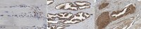
Evaluated by Immunohistochemistry in human prostate.
Immunohistochemistry Analysis: A 1:50 dilution of this antibody detected Utrophin A in human prostate tissue.
Usage Statement
Unless otherwise stated in our catalog or other company documentation accompanying the product(s), our products are intended for research use only and are not to be used for any other purpose, which includes but is not limited to, unauthorized commercial uses, in vitro diagnostic uses, ex vivo or in vivo therapeutic uses or any type of consumption or application to humans or animals.